
11-05-2026 12:32
 Bernard CLESSE
Pourriez-vous m'aider à identifier cette héloti
13-05-2026 15:26
 François Freléchoux
Bonjour,Voici une récolte faite il y a quelques j
12-05-2026 15:41
 Nicolas VAN VOOREN
Dear Ascolovers, especially interested in Pezizale
13-05-2026 12:05
 Thierry Blondelle
Bonjour à tous,J'aimerais avoir confirmation de c
10-05-2026 23:17
 Andreas Gminder
Hello,today we found in a moist steep decidous for
28-04-2026 20:07
 Lothar Krieglsteiner
... on twig in the air at standing Ceratonia siliq
27-04-2026 20:52
Lothar Krieglsteiner
Found on hanging tiwg of Olea europaea in dried-ou
11-05-2026 20:22
Lothar Krieglsteiner
on attached twig of standing Ficus caricaquite uns
29-04-2026 10:44
Lothar Krieglsteiner
growing at moist, drying-out soil at the side of a
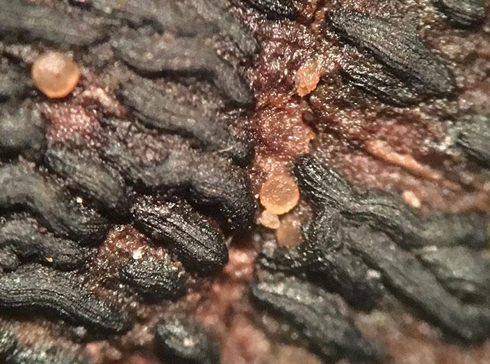
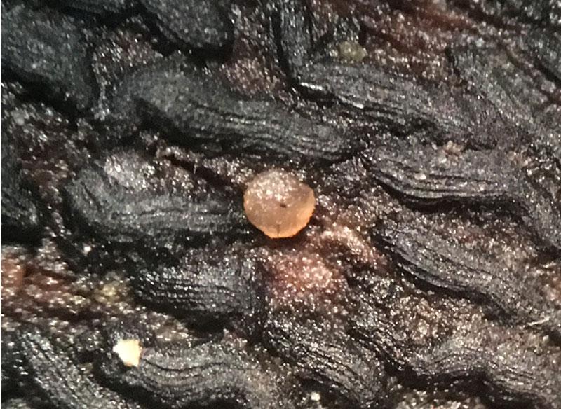
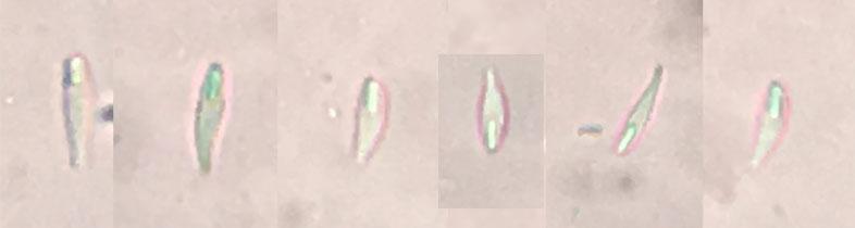
In New York City, Inwood Hill Park. These tiny (.2-.4mm) apothecia were growing on a branch among Hysterobrevium mori. Orange, sessile, disc shaped. The spores are fusoid, narrowing to a point at one end, some curved, most with a long guttule at the wide end, 9-12µm by 2-3µm. The asci I measured were around 31-36µm by 5-6µm. Any help would be appreciated.
Thanks!


this is a great collection, congratulation! Drought-tolerant Orbilias are only sparsely collected so far in the eastern parts of North America, so this seems to be a new record. But I am not sure with the species. This would need more characters to see:
- Please have a look at the ascus apex, especially of more immature asci, and in the dead state: Is the apex only slightly truncate and has it an apical wall thickening?
- Check the excipular cells in the living state for inclusions (globose or variously shaped "crystalloid" ones). Either in a squash mount by external view (don't apply any pressure!) or in a median section.
- Check the paraphyses and their covering exudate.
This sample could belong in the vicinity of Orbilia hesperidea, which is common in the mediterranean area of Europe and which we found in Texas and Arizona. Since it does not occur in temperate Europe, it would be a surprise to see it in New York.
Zotto
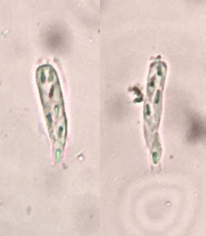
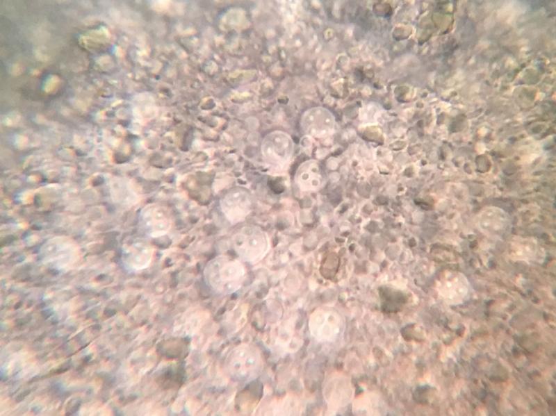
Thank you for your response. I am including a few more pictures and hopefully they will provide the necessary details that you outlined. Here you'll see asci mounted in Lugol's including some immature ones (the two on the far right). I think that shows thickening of the apical wall. An image of the paraphyses (also in Lugol's). Perhaps it shows the exudate you referenced. And finally an external view of the exipular cells in water. Unfortunately I am running out of fruiting bodies, so if i failed to provide the necessary evidence I may not be able to do more.
Again thanks!
Ethan
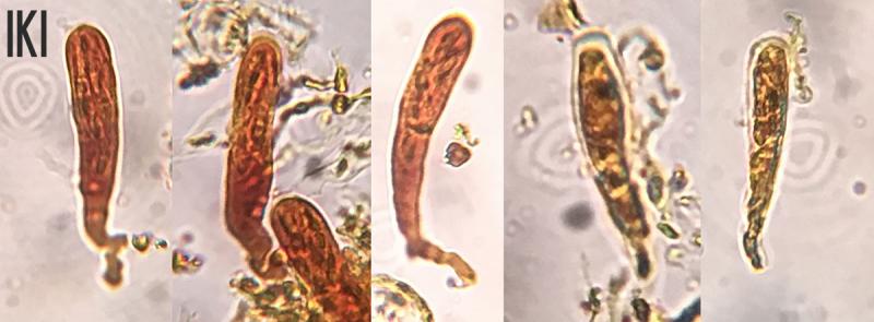
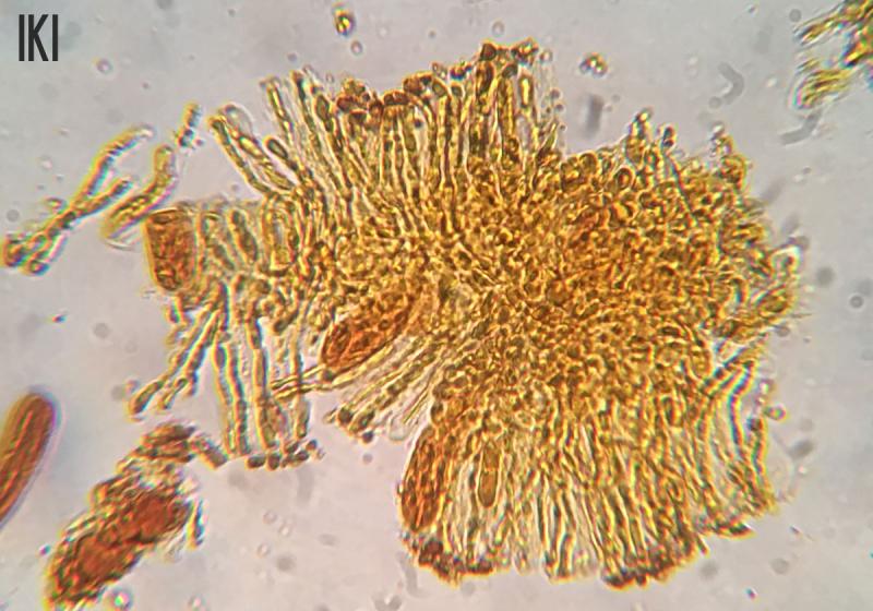
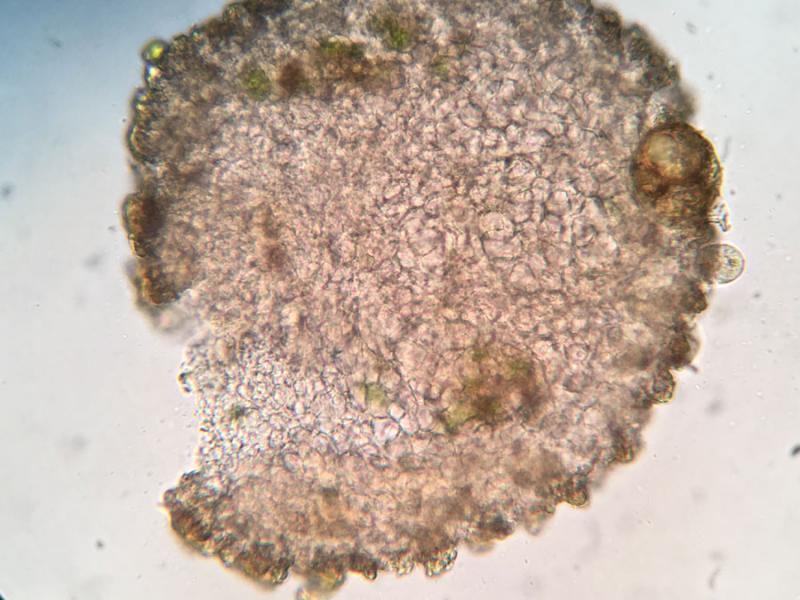
this is helpful, I see the apical wall thickening of the asci and the exudate on the paraphyses. I should have shown you in detail the cell inclusions, because you should have applied oil immersion to see them. Your squash mount is very good, only the resolution of the photo is not enough.
Attached some examples for O. hesperidea.
with SCBs: O. hesperidea
without SCBs: O. montigena
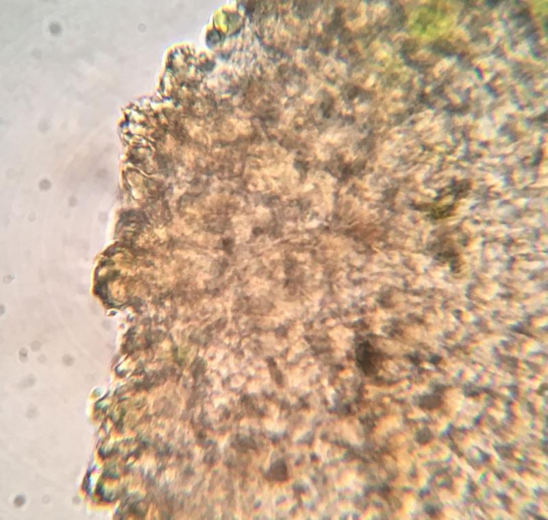
The problem is that I see on your excipular photo distinct small teeth at the margin which might be composed of glassy processes. Such processes are unknown in either of these species here in Europe. Besides, O. hesperidea is a mediterranean species, O. montigena is distinctly montane in central Europe. O. montigena is strictly on angiosperm wood.
What substrate could it be? I assume it is an angiosperm. Was the branch fallen to the ground but not in a moist environment?
Since the site is lowland and the dentate margin unusual, this may well represent an undescribed species unknown to me.
If you tell me the collection date, I could mention this sample in the monograph.
Zotto
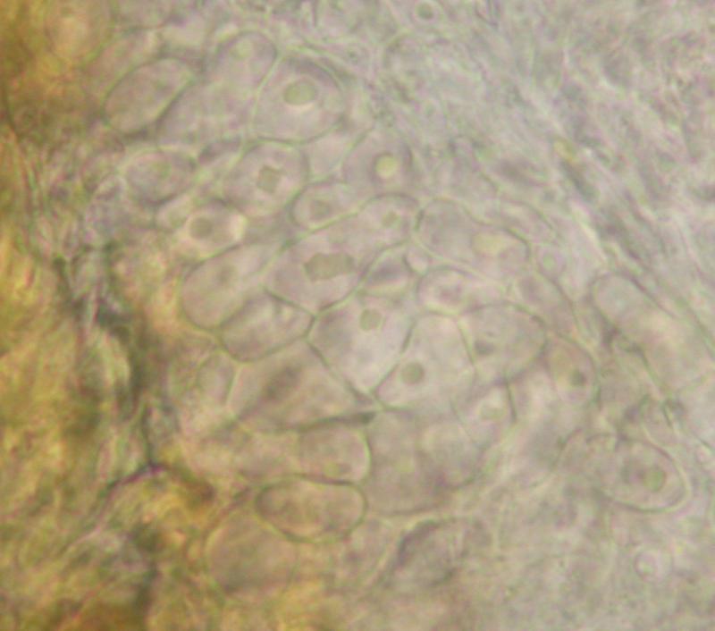
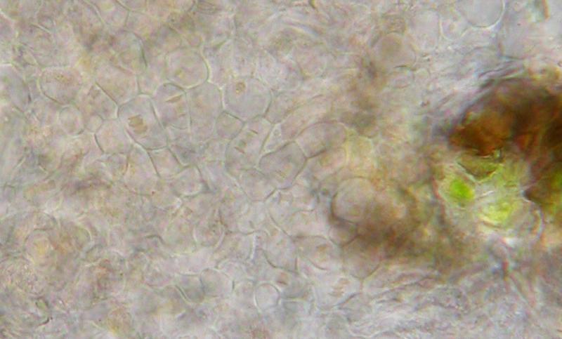
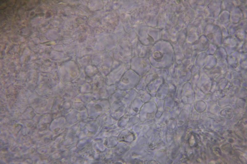
In this photo I have attempted to get closer and perhaps you will recognize inclusions in the exipular cells. I am not certain if that is what is represented by the four spots which appear in many of the cells in the center.
I am also uploading a photo showing the dentate margin.
The collection was made Monday, September 4, 2017 in Inwood Hill Park, the area at the northern tip of Manhattan, somewhere around 40.869759 -73.924269. It was growing on or among Hysterobrevium mori, which I identified using the key in E.W.A. Boehm et. al. "A molecular phylogenetic reappraisal of the Hysteriaceae, Mytilinidiaceae and Gloniaceae (Pleosporomycetidae, Dothideomycetes) with keys to world species" in Studies in Mycology 64: 49–83. 2009. The substrate is angiosperm. The trees in the park are mostly Quercus, Fagus, Liriodendron, Acer among others. The branch was on the ground in a fairly dry area. Unfortunately there are very few fruiting bodies left on the stick, otherwise I would certainly offer to send the collection to you. I don't think it would be useful to you. When I return to Inwood Hill I will look for it again.
Regards,
Ethan
Thanks also for the collection data. Quercus you can easily recognise in a cross section with a hand lens by the large pores arranged in rings, and Fagus by the broad radial rays. No problem if the wood remains undetermined.
I assume you do not keep the sample in a herbarium, do you? I also think it is better for me not to study the remains of this, but if you have anything left you could do another try on the excipulum, just as your previous phto but with oil immersoin.
Zotto