
11-05-2026 12:32
 Bernard CLESSE
Pourriez-vous m'aider à identifier cette héloti
13-05-2026 15:26
 François Freléchoux
Bonjour,Voici une récolte faite il y a quelques j
12-05-2026 15:41
 Nicolas VAN VOOREN
Dear Ascolovers, especially interested in Pezizale
13-05-2026 12:05
 Thierry Blondelle
Bonjour à tous,J'aimerais avoir confirmation de c
10-05-2026 23:17
 Andreas Gminder
Hello,today we found in a moist steep decidous for
28-04-2026 20:07
 Lothar Krieglsteiner
... on twig in the air at standing Ceratonia siliq
27-04-2026 20:52
Lothar Krieglsteiner
Found on hanging tiwg of Olea europaea in dried-ou
11-05-2026 20:22
Lothar Krieglsteiner
on attached twig of standing Ficus caricaquite uns
29-04-2026 10:44
Lothar Krieglsteiner
growing at moist, drying-out soil at the side of a
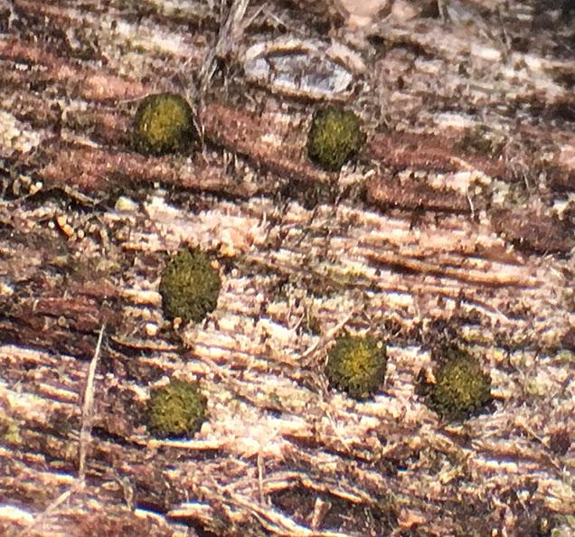
Berkleasmium conglobatum (?)
Ethan Crenson,
11-08-2017 17:19
Hans-Otto Baral,
11-08-2017 19:14

Re : Berkleasmium conglobatum (?)
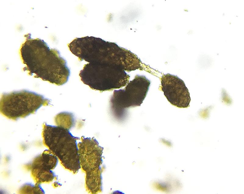
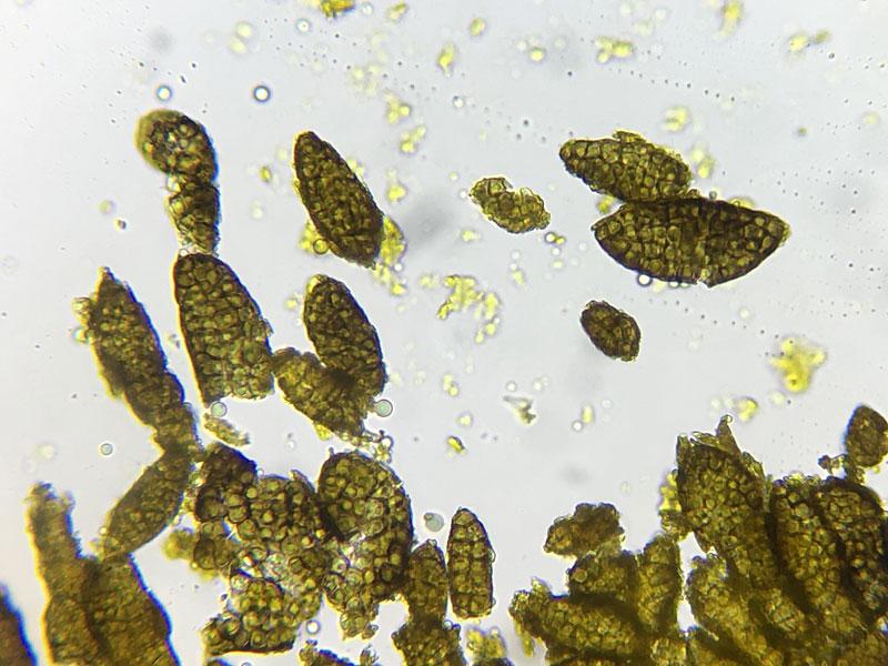
I have seen a similar fungus on dead wood of Acacia in arid Australia, but the conidia were max. 30 µm long. I noticed in this species a strong ionomidotic reaction of the conidia in KOH (orange stain extruding in the medium). Did you test that?
Zotto
Zotto
Jason Karakehian,
11-08-2017 19:50

Re : Berkleasmium conglobatum (?)
Hi Ethan, I posted this species to our Facebook group in June and I just sent you a message with the link to that post. Here is a link to my post in Mycoportal:
http://mycoportal.org/portal/collections/individual/index.php?occid=4622329
I think your determination is correct. The conidia seem to darken in age to nearly opaque black. The farinaceous or flaky condition of the surface of the conidia is consistent with my observations. Also, you will see nearly black sporodochia in a collection and also these yellow-green sporodochia. These are younger sporodochia that have had the tops rubbed away and you see this yellow tissue (hyphae and conidiogenous cells) beneath. Best - Jason