
11-05-2026 12:32
 Bernard CLESSE
Pourriez-vous m'aider à identifier cette héloti
13-05-2026 15:26
 François Freléchoux
Bonjour,Voici une récolte faite il y a quelques j
12-05-2026 15:41
 Nicolas VAN VOOREN
Dear Ascolovers, especially interested in Pezizale
13-05-2026 12:05
 Thierry Blondelle
Bonjour à tous,J'aimerais avoir confirmation de c
10-05-2026 23:17
 Andreas Gminder
Hello,today we found in a moist steep decidous for
28-04-2026 20:07
 Lothar Krieglsteiner
... on twig in the air at standing Ceratonia siliq
27-04-2026 20:52
Lothar Krieglsteiner
Found on hanging tiwg of Olea europaea in dried-ou
11-05-2026 20:22
Lothar Krieglsteiner
on attached twig of standing Ficus caricaquite uns
29-04-2026 10:44
Lothar Krieglsteiner
growing at moist, drying-out soil at the side of a
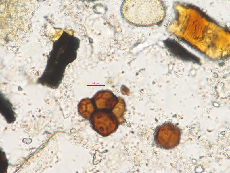
Fungal spores?
Renée Enevold,
21-02-2017 16:26
Thomas Læssøe,
22-02-2017 09:32
Re : Fungal spores?
maybe it would be good to tell where you got the spore from and how it was prepared. I think any wild guess would be welcome :-)
Alain GARDIENNET,
22-02-2017 09:37

Re : Fungal spores?
Yes, Thomas. "One fungus = one name", but : "one spore = one fungus" is not possible, neither "one spore = one name" is possible, logically :)
Dartanha Soares,
22-02-2017 11:41

Re : Fungal spores?
In a perfect world, where guesses become certainties, the "spore" will probably be a pollen grain.
That is my "best guess"
That is my "best guess"
Chris Yeates,
22-02-2017 12:43

Re : Fungal spores?
I too would vote for pollen . . .
Jason Karakehian,
22-02-2017 16:02

Re : Fungal spores?
Maybe it's a bulbil, or bulbil-like conidium. If you think so then try Genera of Hyphomycetes (2011) there are a few pages toward the end of the plates section that treat these. If you have more information (and more than one spore) maybe you can have a go at a determination.
Renée Enevold,
03-03-2017 11:46
Re : Fungal spores?
Thank you very much, Jason. I have had a look in Genera of Hyphomycetes and at the Glomerulomyces fibulosus. Do you think I am on the right track?
Dartanha Soares,
03-03-2017 12:17
Re : Fungal spores?
Glomerulomyces is an hyaline fungus, additionally each cell has only a single pore, your "spore" has several pores in each cell. As I said before, this is more likely to be a pollen grain. It looks like a tetrad, similar to those of Typha, members of Ericaceae, etc.
Renée Enevold,
10-03-2017 12:39
Re : Fungal spores?
No, I am afraid these are not pollen garins, the wall is completely different. These spores are also seen in clumps with more than four cells and often I find single cells. The wall might have been colored by the preparation treatment, but in this case treatment was very mild, only a little KOH and HCl was added, so I assume that it would not color the wall. Could it be any other "bulbil"?
Jason Karakehian,
10-03-2017 13:16
Re : Fungal spores?
Hi Renee, please upload more photos if you have them. In my very limited experience with these, I would think that we may possibly see a hypha attached to one of these structures (if they are fungal). This would have been the point of attachment where this cell (or group of cells) developed from a "conidiogenous" cell. In short - look for any structures that might tell us something about its development, and upload those for us to look at. Also, any measurements, etc. Also, it is always best to mount in water first and make observations, then add KOH or anything else. Some fungi have strong reactions even with a 2% concentration of KOH, turning their walls darker colors of brown or even green in the case, for example, of the mycorrhizal basidiomycete genus Tomentella. You will want to make note of any color changes in different mountants, but use water as your "base line". Best - Jason
PS - Also, does the literature that is specific to your line of work discuss how to work with fungal samples at all? One would think that the chances of finding pollen grains in a sediment sample might be higher than finding more than one fungal spore of the same species in that sample - but I don't know?
PS - Also, does the literature that is specific to your line of work discuss how to work with fungal samples at all? One would think that the chances of finding pollen grains in a sediment sample might be higher than finding more than one fungal spore of the same species in that sample - but I don't know?