
27-04-2026 18:48
Tony MoverleyCollected 23rd April 2026, Norfolk, EnglandSwarms
27-04-2026 20:52
 Lothar Krieglsteiner
Found on hanging tiwg of Olea europaea in dried-ou
28-04-2026 22:51
 Bernard CLESSE
Bonsoir à toutes et tous,Pourriez-vous m'aider à
29-04-2026 08:01
Lothar Krieglsteiner
... on twig attached to small tree of Citrus auran
29-04-2026 10:44
Lothar Krieglsteiner
growing at moist, drying-out soil at the side of a
28-04-2026 20:33
Vitus SchäfftleinHello, I found Trochila ilicina on Ilex aquifoliu

28-04-2026 21:50
 Pablo Sandoval
Hola a todos,Espero se encuentren bien. Hace mucho
27-04-2026 18:05
Lothar Krieglsteiner
... still attached at standing tree. The green con
28-04-2026 20:07
Lothar Krieglsteiner
... on twig in the air at standing Ceratonia siliq
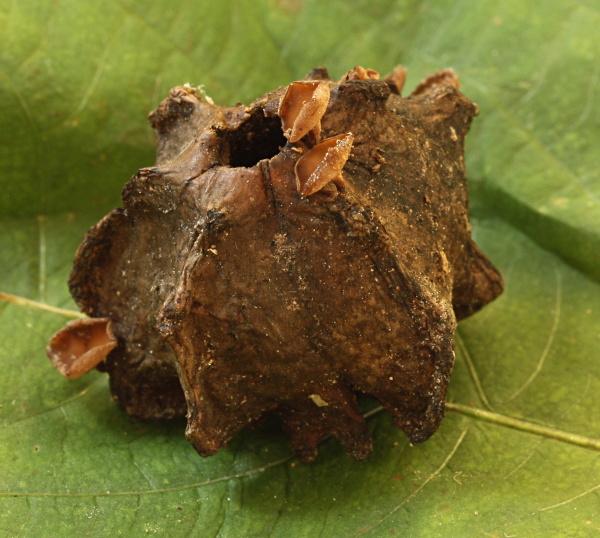
Ciboria americana?
Viktorie Halasu,
24-08-2016 17:53
 Hello, I'd like to ask for your help with this inoperculate on an uncommon host.
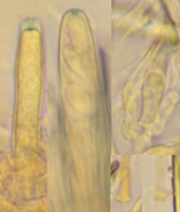
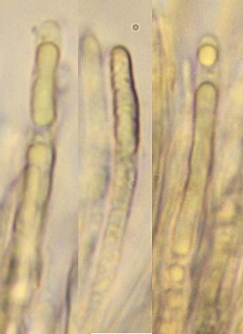
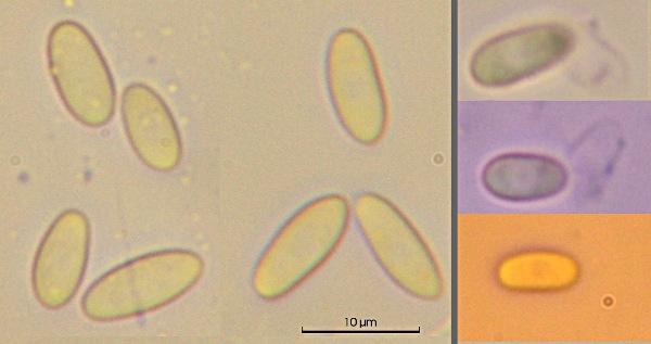
 Apotecia up to 4 mm diam, hymenium cinnamon brown, outside lighter, with short stipe, growing on galls of Andricus cf. quercuscalicis, in Czech Republic. Unfortunately I didn't notice, what kinds of oaks were growing nearby, but this insect usually attacks acorns of Quercus robur/petrae/rubra.
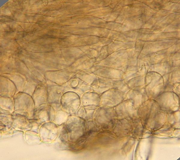
Ectal excipulum of thin t. globulosa, some cells contain vacuoles that stain light azure blue in CRB.
Medulla of interwoven hyphae running mostly parallel to the surface.
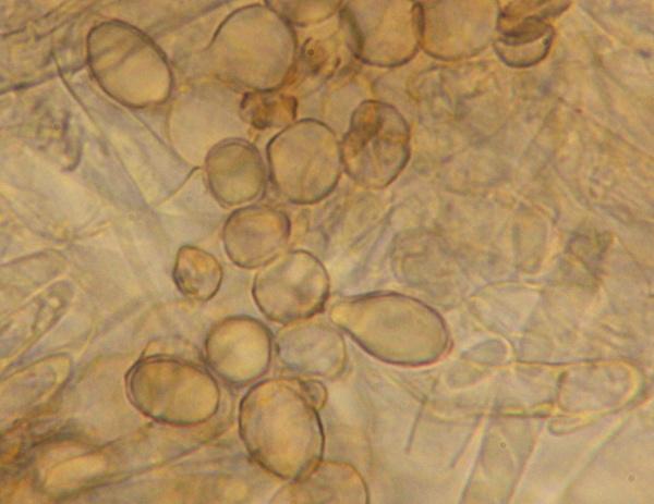
Asci 8-spored, IKI+ (b), croziers+.
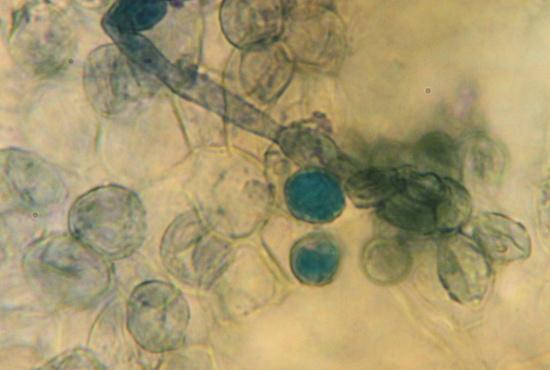
Paraphyses contain long VB in upper part, those also stain light blue in CRB.
Spores (fusoid-)ellipsoid, eguttulate, uninucleate, non-septate, with a loosening gel sheath, 8,5-9,8 (11) × 4,4-4,9 um (average 9,3 × 4,6 um), Q = 1,8-2-2,3 (sporeprint in water).
Is it Ciboria americana? The spores seem a bit too short.
Thank you in advance.
Hans-Otto Baral,
24-08-2016 18:39

Re : Ciboria americana?
Very interesting collection! I measure from your scale slightly narrower spores: 7.7-10.7 x 4-4.5 µm.
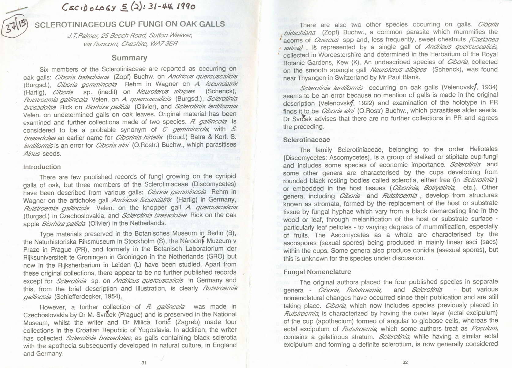
There is a Rutstoemia gallincola on Andricus quercuscalicis redescribed by Palmer 1990, which should better belong in Ciboria and has spores 6,5-11 x 2,7-4 µm. Possibly the shrinking effect explains this difference.
I attach the article.
Zotto
There is a Rutstoemia gallincola on Andricus quercuscalicis redescribed by Palmer 1990, which should better belong in Ciboria and has spores 6,5-11 x 2,7-4 µm. Possibly the shrinking effect explains this difference.
I attach the article.
Zotto
-
 Palmer-1990-Sclerotiniaceae-on-oak-galls-1-0002.jpg
 -
Palmer-1990-Sclerotiniaceae-on-oak-galls-2-0002.jpg
-
Palmer-1990-Sclerotiniaceae-on-oak-galls-3-0002.jpg
-
Palmer-1990-Sclerotiniaceae-on-oak-galls-4-0002.jpg
-
Palmer-1990-Sclerotiniaceae-on-oak-galls-5-0002.jpg
-
Palmer-1990-Sclerotiniaceae-on-oak-galls-6-0002.jpg
-
Palmer-1990-Sclerotiniaceae-on-oak-galls-7-0002.jpg
 Palmer-1990-Sclerotiniaceae-on-oak-galls-1-0002.jpg
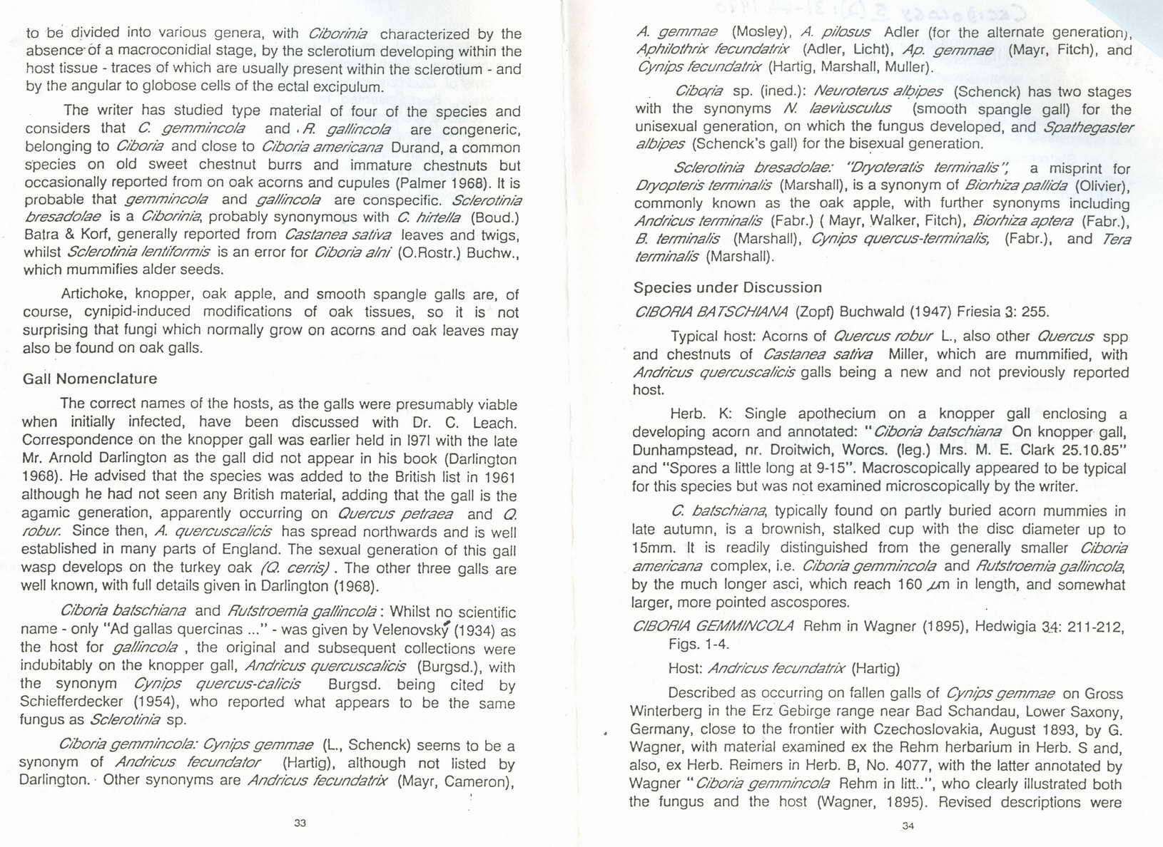 Palmer-1990-Sclerotiniaceae-on-oak-galls-2-0002.jpg
 Palmer-1990-Sclerotiniaceae-on-oak-galls-3-0002.jpg
 Palmer-1990-Sclerotiniaceae-on-oak-galls-4-0002.jpg
 Palmer-1990-Sclerotiniaceae-on-oak-galls-5-0002.jpg
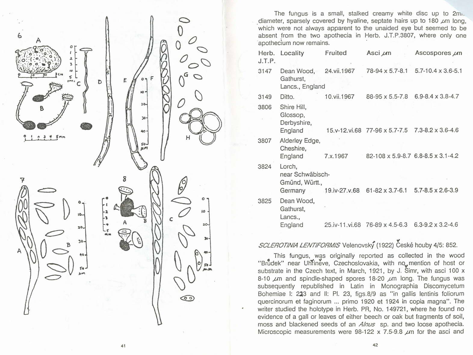 Palmer-1990-Sclerotiniaceae-on-oak-galls-6-0002.jpg
 Palmer-1990-Sclerotiniaceae-on-oak-galls-7-0002.jpg
Viktorie Halasu,
24-08-2016 18:50
Re : Ciboria americana?
Thank you very much, Mr. Baral. I should have thought that there are no easy answers with inoperculates! :) I'm not at home right now, but next week I'll measure again the spore size from exsiccate.
Hans-Otto Baral,
24-08-2016 20:32
Re : Ciboria americana?
This is a good idea, and please take a reagent that kills the spores (KOH, Melzer or cotton blue-lactophenol), because they surely survive some weeks in the dry state.
And please keep this material, it would be great at some time to clarify its molecular relationship, because Ciboria is heterogeneous.
And please keep this material, it would be great at some time to clarify its molecular relationship, because Ciboria is heterogeneous.
Viktorie Halasu,
29-08-2016 15:23
Re : Ciboria americana?
Good afternoon, Mr. Baral,
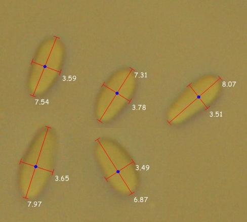
previously, the sporeprint contained like 50:50 of bigger and smaller spores (cca 10 and 7-8 um long, respectively), they looked like two separate groups. I thought the smaller ones were immature and maybe measured more of the longer ones. Another measuring (from the same sporeprint in water) where I included more of the smaller ones, gave (6,4) 7-9,5 (11) × (3,3) 3,4-4,7 (4,9) um, avg. 8,4 × 4,1 um.
In exsiccate there's only a few of the longer spores and they're not so clearly differentiated. Dimensions in Melzer fit the R. gallincola better: (6,5) 7-8,7 (9,3) × (3,1) 3,2-3,7 (3,8) um, avg. 7,8 × 3,4 um. Asci are not blueing in MLZ (or so faintly, that I can't see it for sure with my microscope), neither any part of excipulum.
I'll send you the collection data via e-mail.
Just to make sure - is this the correct way to measure the spores? (It's a screenshot from Piximétre, photos taken with 40× objective.)
Hans-Otto Baral,
29-08-2016 21:00
Re : Ciboria americana?
I personally do not like much this method of measuring. But your photos are not really sharp, and I would tentatively put the scale a bit shorter.
Good that the dead spores match this fungus. Nevertheless we must be cautious whether this is a species different from C. americana, or it was mainly erected because of the strange substrate.
Good that the dead spores match this fungus. Nevertheless we must be cautious whether this is a species different from C. americana, or it was mainly erected because of the strange substrate.