27-04-2026 20:52
 Lothar Krieglsteiner
Found on hanging tiwg of Olea europaea in dried-ou
27-04-2026 18:48
Tony MoverleyCollected 23rd April 2026, Norfolk, EnglandSwarms
27-04-2026 17:41
Lothar Krieglsteiner
.. Algarve, same leaf than the last post. The con
27-04-2026 18:05
Lothar Krieglsteiner
... still attached at standing tree. The green con
27-04-2026 17:16
Lothar Krieglsteiner
.. Algarve, moist lying.The conidiomata look like
27-04-2026 12:54
Steve ClementsBonjour. Ce petit champignon blanc résupiné et
27-04-2026 09:59
Pauline. PennaBonjour Can anyone advise me on these pycnidia fo

22-04-2026 20:54
 Enrique Rubio
Hi to everybody.This Pyrenopeziza grew in moist le
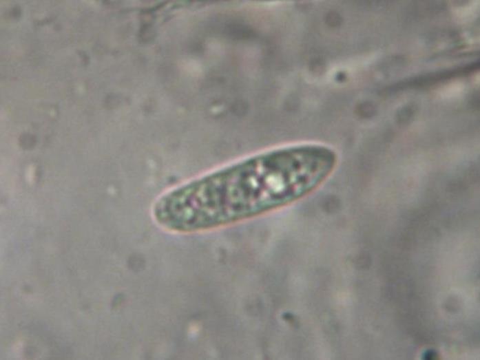
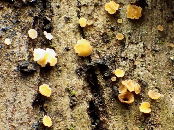
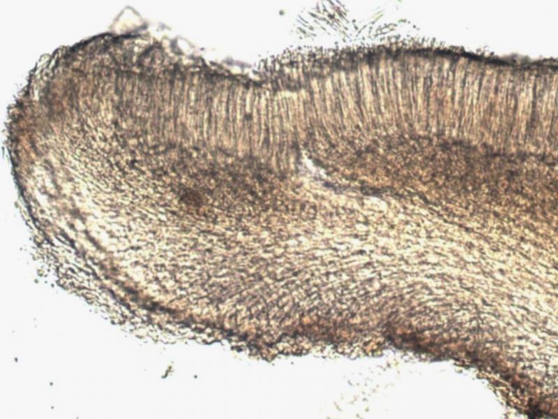
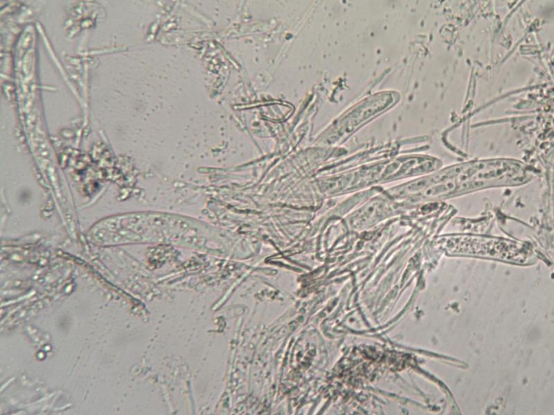
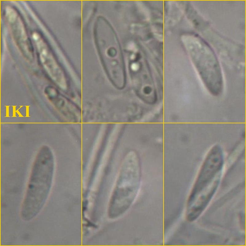
I found this cups growing on the bark of Eucalyptus. It seems to be Mollisia or close. However, the spores are a bit to large. I got the following dimensions for them:
(9.7) 9.8 - 14.1 (14.6) × (2.2) 2.4 - 3.7 (3.9) µm
Q = (2.8) 3.1 - 4.4 (4.6) ; N = 23
Me = 11.9 × 3.3 µm ; Qe = 3.7 .
I attach a set of photos with its features.
I would like to have some help for its classification.
Thanks in advance,
zaca

Hello,
the spores wouldn't rule out Mollisia, but all the rest doesn't fit.
I think this is a Hymenoscyphus or a related genus.
best regards,
Andreas
Let's move the target genus.
Best regards,
zaca

Zotto
Hello,
may be it's worth comparing with Hymenoscyphus phialea.
best regards,
Andreas

Bonjour,
I would suggest a comparison with Bisporella subpallida, at least the macro would fit and also the excipulum ?
Amitiés
Michel
Many thanks for your comments, opinions and proposals.
I'm a bit confused by now: I could not find any information about Hymenoscyphus phialea, suggested by Andreas, and regarding Bisporella subpallida, mentioned by Michel and Zotto, that species has spores much smaller than my specimen, despite of the similar macro appearance, as I could see in several places. I checked again the dimensions of the spores.
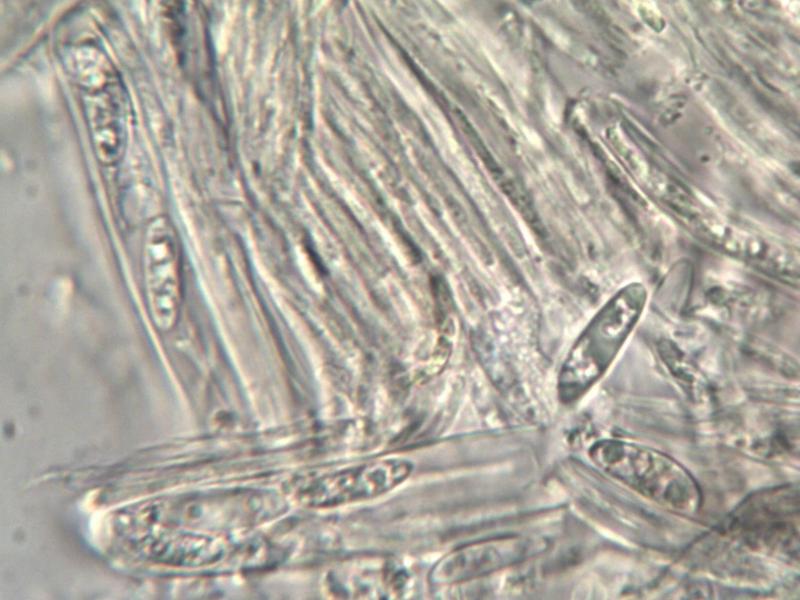
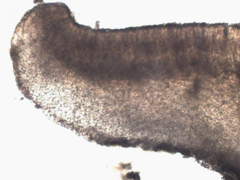
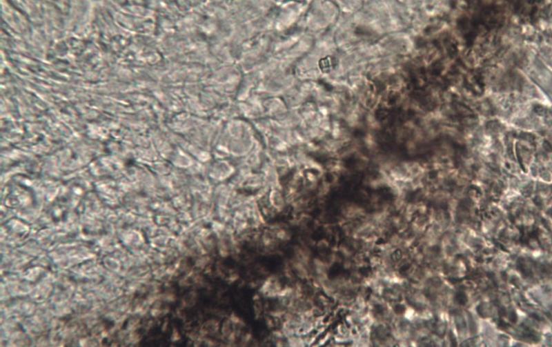
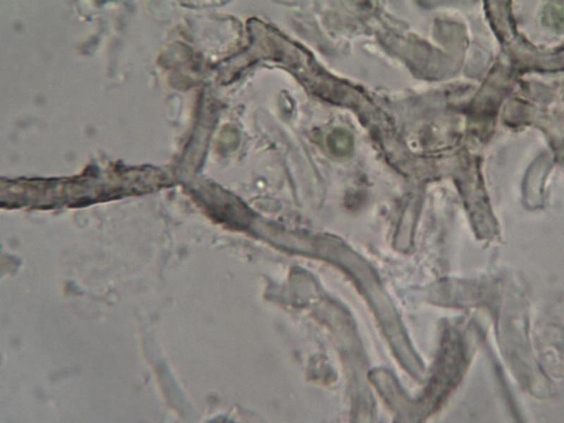
I take the oportunity to upload two photos, one of the ectal exciple and the other with some strange hyphae seeming incrusted.
Regards,
zaca
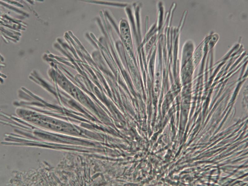
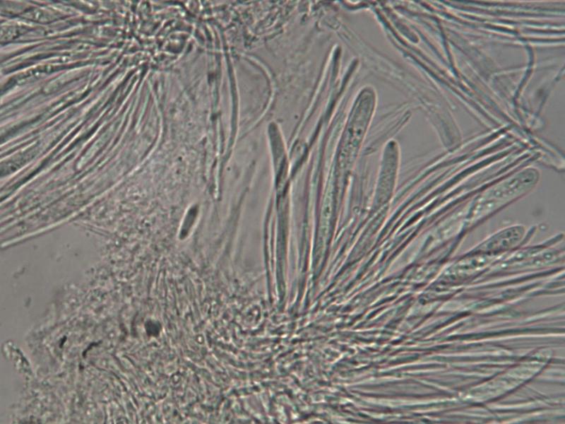
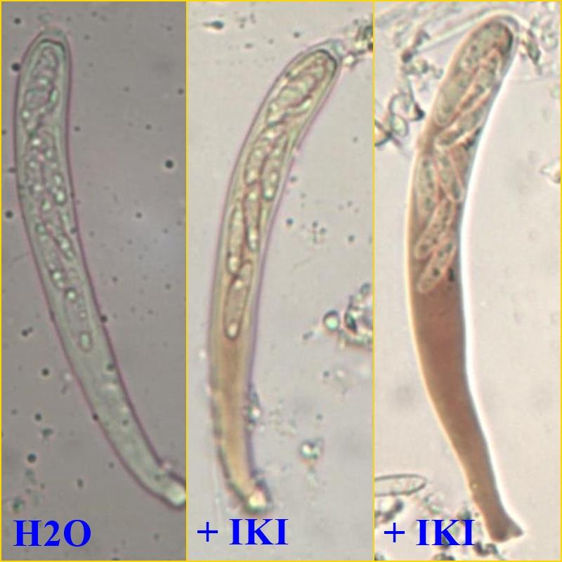
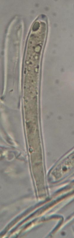
The asci look amyloid, do they? The shape of ring would really be helpful.
The correct name for Andreas' suggestion is Helotium phiala, but that species has inamyloid asci.
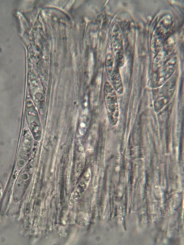
I will repeat the microscopy and if something new I will post it here. As far as I can see from the previous micro, it is difficult to classify the asci as amyloid since there is a very diffuse bluish tone at the apex and that's all, but if you see the photo in water the appex already seems a bit blue. I also took a photo, that I upload now, of the Lugol reaction in a mount with KOH and that gave blue apex.
Best regards, zaca
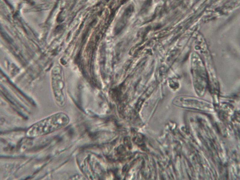
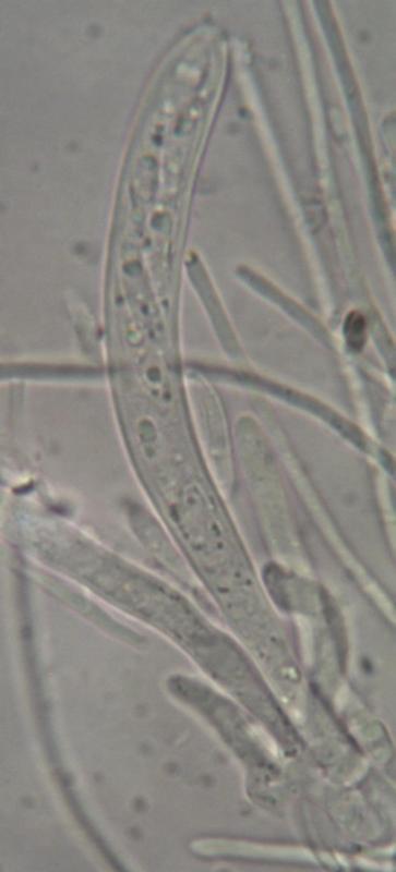
If you find the fungus again, please make preparations in water with only slight pressure on the cover slip. maybe we can then proceed in finding a genus.
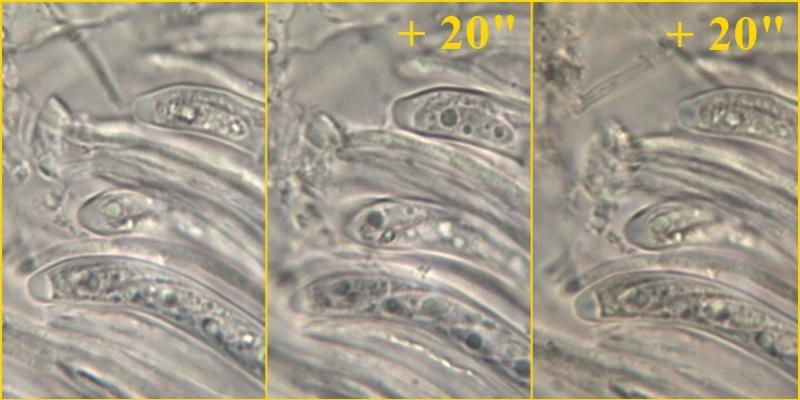
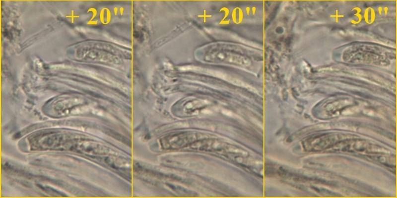
Following the recommendations of Zotto I remade the microscopic observation, using a mount in water to observe the spores; After joined IKI and analyzed the evolution of the reaction, taking photos at regular intervals. As expected from the previous observation, the bluish apex reaction of asci is very discrete. I attach some photos, hoping that these can be useful.