05-05-2026 22:40
Gernot FriebesHi,I believe this is a Plagiostoma growing on a Sa
06-05-2026 11:25
 Castillo Joseba
Me mandan el material seco de Galicia (España) re
06-05-2026 17:23
Thomas Læssøehttps://svampe.databasen.org/observations/10594257
28-04-2026 20:07
 Lothar Krieglsteiner
... on twig in the air at standing Ceratonia siliq
04-05-2026 18:13
 Stephen Martin Mifsud
ID request for what seems to be a true aquatic fun
04-05-2026 16:39
Stephen Martin Mifsud
ID request: This specimen was collected in Malta o
28-07-2011 18:31
 Alex Akulov
Dear FriendsToday I made the pdf file of Velenovsk
04-05-2026 09:50
Castillo Joseba
Me mandan el material seco de Galicia,(España) re

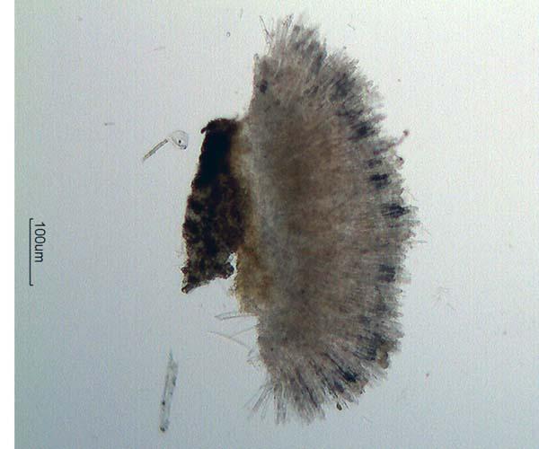
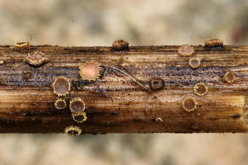
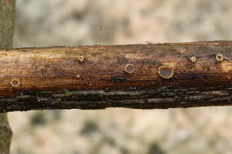
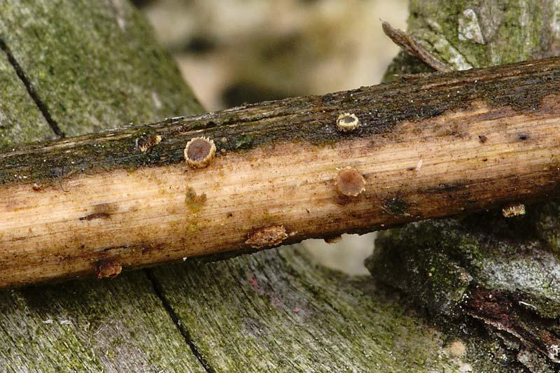
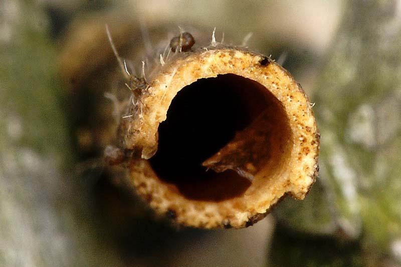
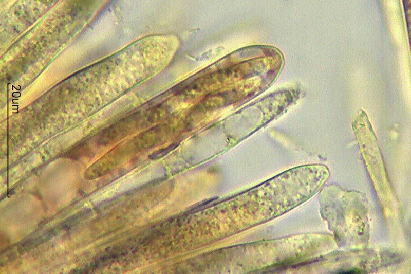
Hola a todos.
Subo unas fotos de un asco que hemos encontrado hoy casi seguro sobre ramitas de hinojo.
Miden hasta 2 mm de diámetro.
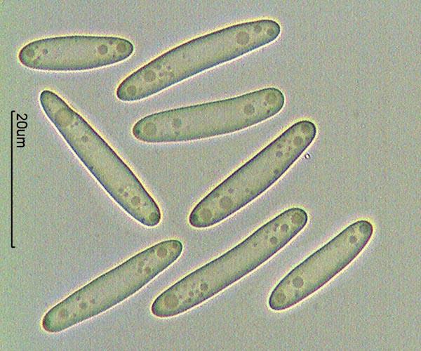
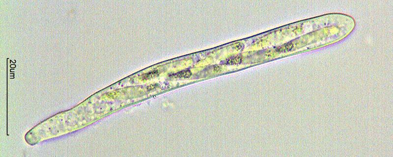
Esporas de 17-27 x 4-5 micras.
Ascas IKI- (no aprecio reacción hemiamiloide) y creo que aporrincas.
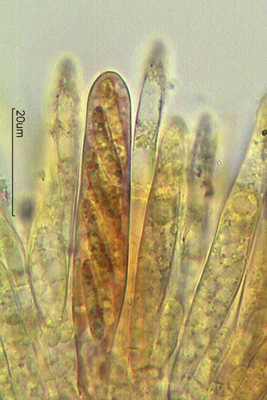
Paráfisis lanceoladas hialinas de perfil rugoso y con presencia de cristales (creo)
¿Qué les parece? Pensé en Trichopeziza "perrotioides" pero tengo dudas.
Gracias por su ayuda.
Rubén
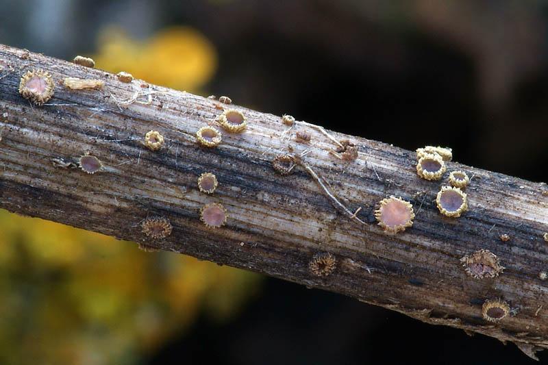
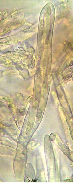
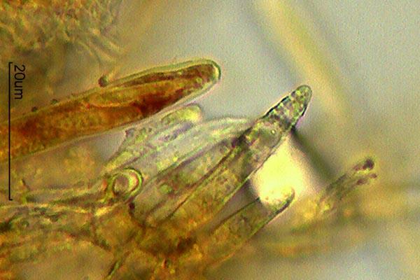
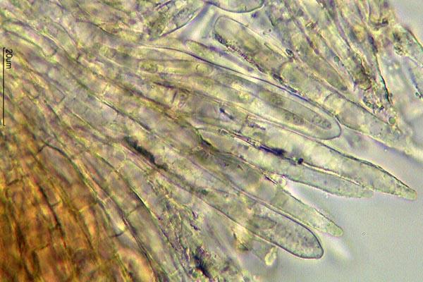


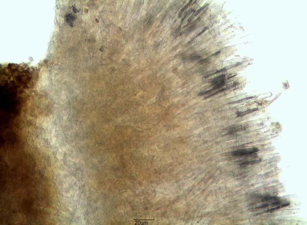
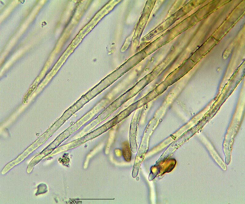
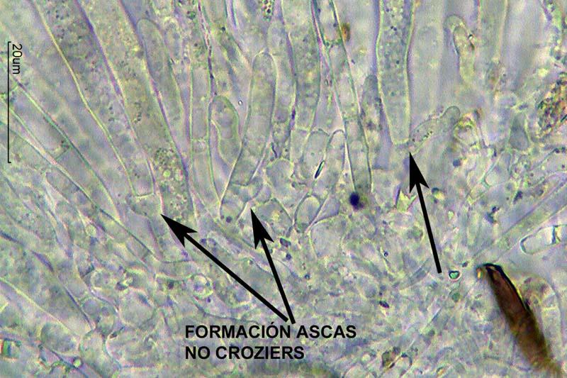
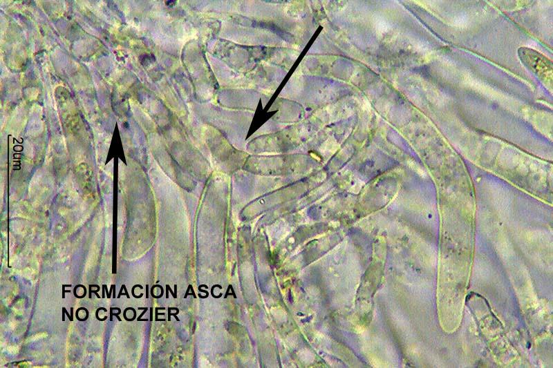
thanks for this wonderful documentation! It is obviously very close to my Trichopeziza "perrotioides", e.g. in the rough paraphyses, but differs in much larger straight spores. Are the hairs not longer than about 100 µm? The asci are obviously inamyloid, but it would be great if you succeed to make a photo of the ascus base (perrotioides is without croziers).
Please keep this sample in your herbarium, I am sure it will be needed in a future study, e.g. for a sequence. Could well be a new species.
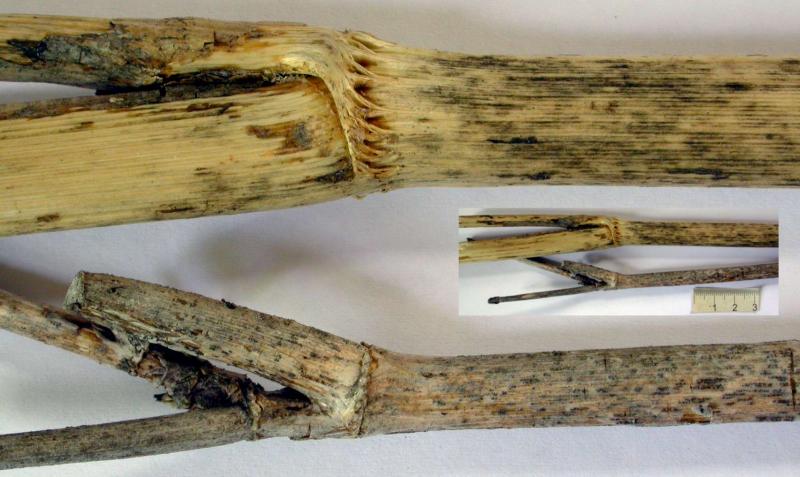
You can try lookin at the anatomy of the stem, I have here photos of Foeniculum.
Zotto
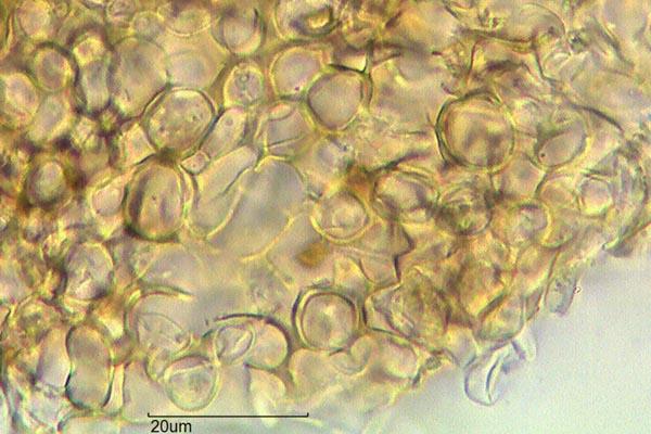
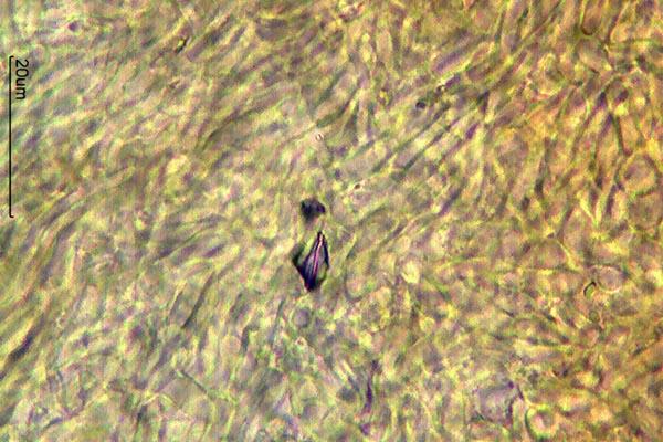
Hola a todos.
Gracias por su ayuda, Zotto. Bonitas fotos! Yo no consigo hacer macro tan bueno de tallo de Foeniculum.
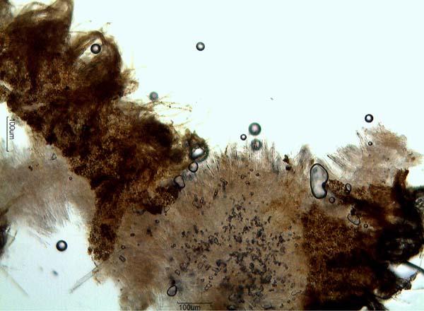
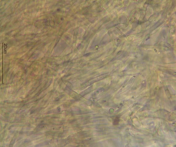
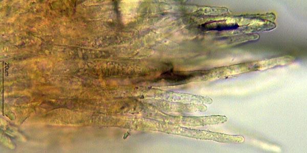
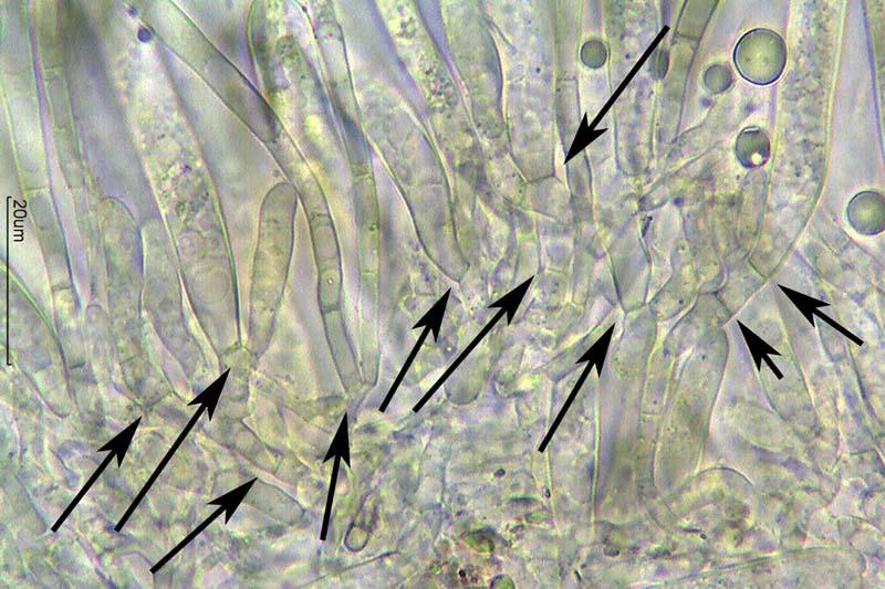
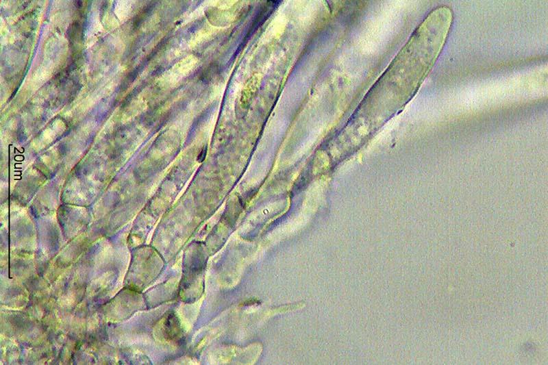
He mirado base de ascas y no he visto croziers. A verces parece que sí por la formación de nueva asca. Pongo fotos.
Los pelos son más grandes, miden aproximadamente hasta 250 micras, pero a veces saco fotos a pequeños para ver la formación.
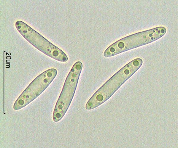
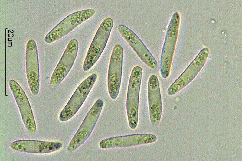
Paso fotos de esporas recién expulsadas del asca y tienen medidas de anchura algo menor (3,3-4,3 micras) y también de asca IKI-.
Saludos
Rubén
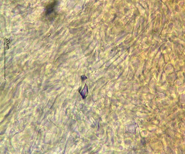
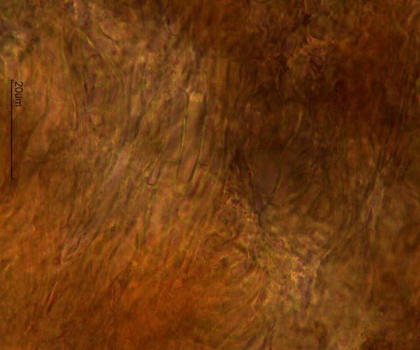
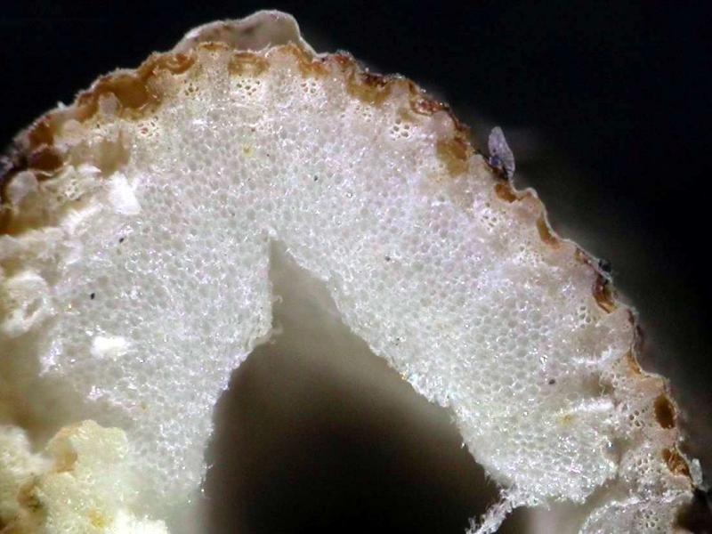
Your stem differs from mine in lacking the thick internal parenchyma. Also I wonder whether there are vascular bundles at different levels, not only in one row. That would be typical of monocots! To clarify this you could try a section viewed under the microscope (100x).
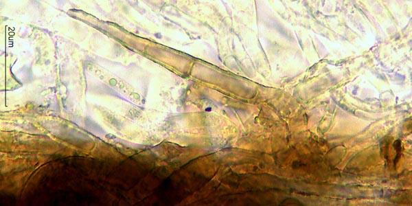
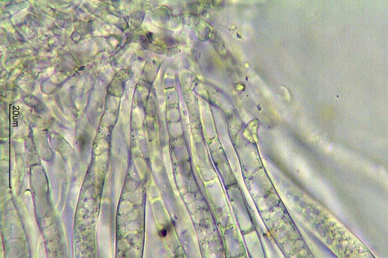
Hola Zotto, gracias por su ayuda.
Ya tengo ejemplares todos secos, pero ayer hice fotos a 100X. Pongo alguna por si sirve.
Saludos
Rubén