
05-05-2026 22:40
Gernot FriebesHi,I believe this is a Plagiostoma growing on a Sa
04-05-2026 18:13
 Stephen Martin Mifsud
ID request for what seems to be a true aquatic fun
04-05-2026 16:39
Stephen Martin Mifsud
ID request: This specimen was collected in Malta o
28-07-2011 18:31
 Alex Akulov
Dear FriendsToday I made the pdf file of Velenovsk
28-04-2026 20:07
 Lothar Krieglsteiner
... on twig in the air at standing Ceratonia siliq
04-05-2026 09:50
 Castillo Joseba
Me mandan el material seco de Galicia,(España) re
02-05-2026 12:42
Alain BRISSARDBonjour à tousJeuidi 30 avril dernier on m'a remi
02-05-2026 13:06
Pauline. PennaBonjour Please can someone help me with this id
01-05-2026 22:45
 Thierry Blondelle
Bonjour à tous, Une récolte sur bouse séchée d
Dicephalospora
Zuzana Sochorová (Egertová),
13-06-2015 21:05
 Hello,
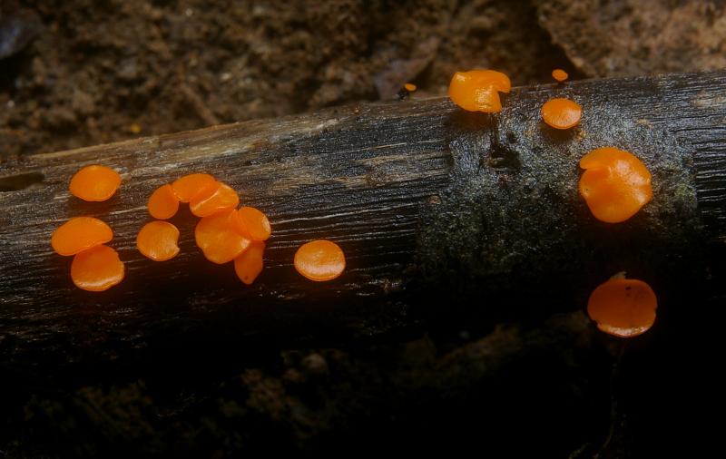
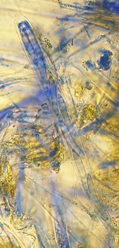
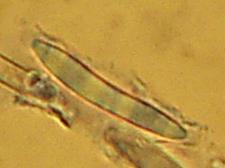
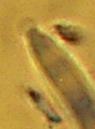
this ascomycete was found in a karst region on Borneo. It should be Dicephalospora (it has round caps on the poles of spores and macroscopically it fits well too). There are two species with IKI+ asci and spores narrower than 8 micrometers in the key by Verkley 2004 - D. rufocornea and D. calochroa. The problem with D. rufocornea is that my spores are twice shorter, for D. calochroa my spores are twice thinner. Could it be another genus? Or could the drying change the spore size so rapidly (I worked with dried material)?
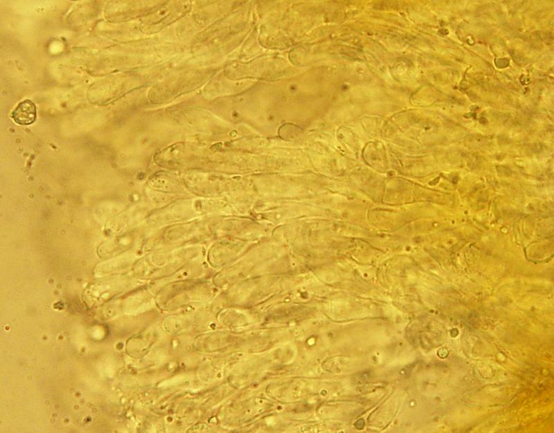
Apothecia orange, with a rounded disc in the youngest fruitbodies, irregularly waved in the elders, 0.5-6 mm broad, flat, very slowly convex or very slightly concave, stipe short, whitish, up to 2 mm long.
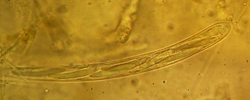
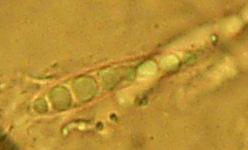
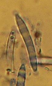
Asci 121-130 x 7-8 micrometers, octosporic, IKI+.
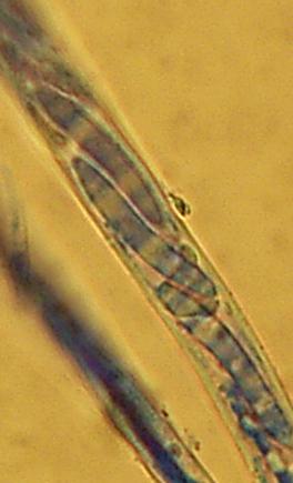
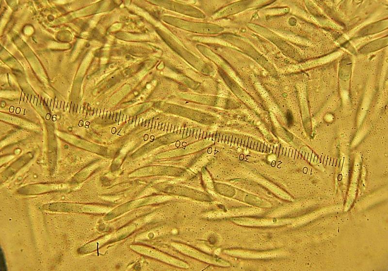
Spores 19.5-24 x 2.5-3.5 micrometers, usually biseriate, smooth, hyaline.
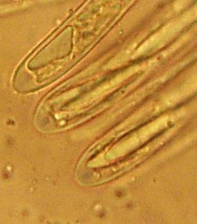
Paraphyses hyaline, 1-1.5 micrometers wide.
Thanks, Zuzana
Hans-Otto Baral,
13-06-2015 23:47

Re : Dicephalospora
Hi Zuzana
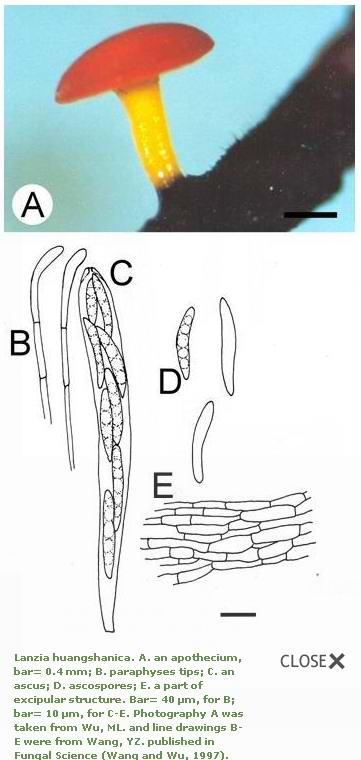
perhaps "Lanzia" huanshanica, though this is perhaps without terminal sporre caps.
Lanzia huangshanica. A. an apothecium, bar= 0.4 mm; B. paraphyses tips; C. an ascus; D. ascospores; E. a part of excipular structure. Bar= 40 µm, for B; bar= 10 µm, for C-E. Photography A was taken from Wu, ML. and line drawings B-E were from Wang, YZ. published in Fungal Science (Wang and Wu, 1997). Author: Lanzia huangshanica W. Y. Zhuang & Korf., Mycosystema 7: 13-17. 1995. Description: Stroma black, substratal. Apothecia 0.3-1.2 mm, discoid, red to orange red when fresh, dark red when dry, stipitate, 0.3-0.7mm high. Receptacle yellow, stalk yellowish orange, black at base. Ectal excipulum of textura prismatica, thick-walled, cells 7-17 × 2.5-4 µm. Medullary excipulum of textura intricata. Asci 8-spored, clavate, apical pore turning blue in Melzer's reagent, 87-97 × 8-9.5 µm. Ascospores, long fusiform, hyaline, 19-25 × 3-4.5 µm, with guttules. Paraphyses 1.8-2.0 µm at base, slightly enlarged and curved at tips, 2-4 µm wide. Specimens: Taiwan. Yilan: Fushan Botanical Garden; alt. 660-750 m, on petiole of broad-leaved trees, 24 Mar. 1994, Wu94-F2-II-L1. 29 Oct. 1995, Wu95-F10-II-L1; 27 June 1996, Wu96-F6-II-L1; on the leaf petioles of Fagaceae sp., 9 Mar. 1994, WAN008; on the leaf petioles of Castanopsis sp., 16 June 1995, WAN169. Habitat: Foliicolous. On the petiole of broad-leaved trees such as Castanopsis sp. and Fagaceae species. Distribution: China, Japan and Taiwan. References: Wang, YZ and Wu, ML. 1997; Zhuang, WY. 1995. Provided: M. L. Wu Note: It is similar to Dicephalospora rufocornea by the orange-red, stipitate apothecia, fusoid ascospores, and excipular structures, but the latter has larger and brighter colored apothecia, asci and ascospores. The ascospores of D. rufocornea have gelatinous collar at poles and the paraphyses are straight not curved at the tips. Zhuang (1995) mentioned the apothecial tissues does not exude pigment in 2% KOH solution, however, small amount of dark red pigment emitting from dry specimens in KOH solution was observed from Taiwan sample. Collections were found all year round at Fushan Botanical Garden, on dead leaf petioles of Fagaceae species.
Zuzana Sochorová (Egertová),
14-06-2015 07:30
Re : Dicephalospora
Thank you, Zotto. The microscopy in this species is much closer to my fungus than in Dicephalosporas in the key (although mine has caps). Macroscopically, it´s not so perfect - my fungus has up to 5x larger disc (and very brightly coloured, I would say), whitish stipe and grows on wood, not on leafs.
Hans-Otto Baral,
14-06-2015 07:44
Re : Dicephalospora
ah o.k., that was on leaves, I overlooked. Of course you have the fungus only in dry state - I suspect the caps are better seen in living state. You don't have free spores?
Zuzana Sochorová (Egertová),
14-06-2015 08:13
Re : Dicephalospora
Yes, only dried, it would be impossible to take microscope to those Bornean conditions... I saw the caps well, but they are not easy to photograph. I will make more photos in the evening, including free spores.
Zuzana Sochorová (Egertová),
14-06-2015 19:20
Re : Dicephalospora
Some more notes:
- few spores are up to 4 micrometers broad
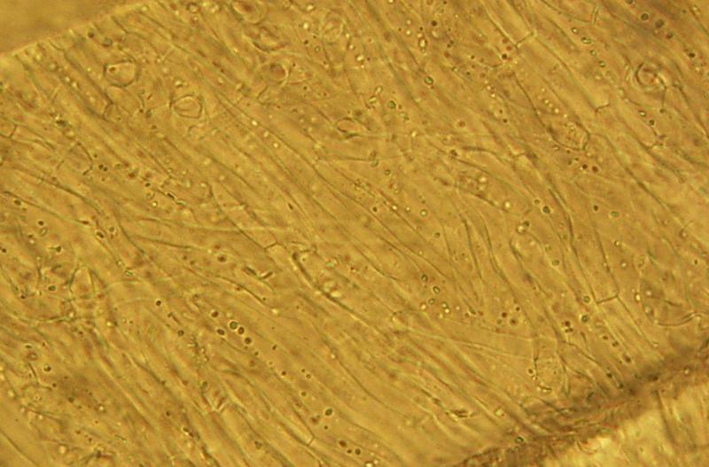
- excipulum med. consists of textura itricata (cells 2.5-4 micrometers broad)
- excipulum ect. consists of textura porrecta (cells 2-6 micrometers broad, with somewhat thicker walls then cells in exc. med.)
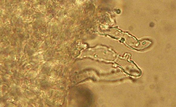
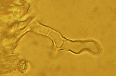
- there are short, thick-walled, septate hairs with small excrescenses on the outer surface (about 3.5 micrometers broad). Macroscopically, the outer surface of the dried apothecia is completely whitish.
- paraphyses are straight
I hope the caps are visible in the pictures.
Zuzana
- few spores are up to 4 micrometers broad
- excipulum med. consists of textura itricata (cells 2.5-4 micrometers broad)
- excipulum ect. consists of textura porrecta (cells 2-6 micrometers broad, with somewhat thicker walls then cells in exc. med.)
- there are short, thick-walled, septate hairs with small excrescenses on the outer surface (about 3.5 micrometers broad). Macroscopically, the outer surface of the dried apothecia is completely whitish.
- paraphyses are straight
I hope the caps are visible in the pictures.
Zuzana
Hans-Otto Baral,
14-06-2015 20:43
Re : Dicephalospora
Yes of course, clearly with apical caps. I have looked at the two species dscribed by Zhuang 1999, but they have also much to wide spores.
Interesting!
Interesting!
Zuzana Sochorová (Egertová),
14-06-2015 21:30
Re : Dicephalospora
Hmm, it´s bad - 4 collections of Dicephalospora from Borneo, and 0% success in determination :-)
There are only D. chrysotricha and D. rufocornea listed in the Checklist of Fungi of Malaysia.
Thanks for help!
There are only D. chrysotricha and D. rufocornea listed in the Checklist of Fungi of Malaysia.
Thanks for help!