05-05-2026 22:40
Gernot FriebesHi,I believe this is a Plagiostoma growing on a Sa
04-05-2026 18:13
 Stephen Martin Mifsud
ID request for what seems to be a true aquatic fun
04-05-2026 16:39
Stephen Martin Mifsud
ID request: This specimen was collected in Malta o
28-07-2011 18:31
 Alex Akulov
Dear FriendsToday I made the pdf file of Velenovsk
28-04-2026 20:07
 Lothar Krieglsteiner
... on twig in the air at standing Ceratonia siliq
04-05-2026 09:50
 Castillo Joseba
Me mandan el material seco de Galicia,(España) re
02-05-2026 12:42
Alain BRISSARDBonjour à tousJeuidi 30 avril dernier on m'a remi
02-05-2026 13:06
Pauline. PennaBonjour Please can someone help me with this id
01-05-2026 22:45
 Thierry Blondelle
Bonjour à tous, Une récolte sur bouse séchée d
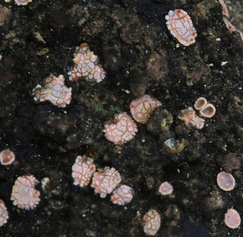
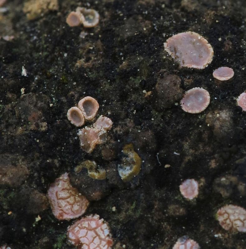
Pink lichen-like asco
Stephen Martin Mifsud,
31-03-2015 09:25
Hi guys, I am attempting a determination on this beautiful pinkish lichen-like ascomycete (saw some small cups, and not greenish so, I am inclined it is a fungus). I will do some micro and keep you posted. It is so beautiful that I could not resist not posting! It was located on limestone-rich damp soil exposed and in full sunshine. (that favours a lichen!)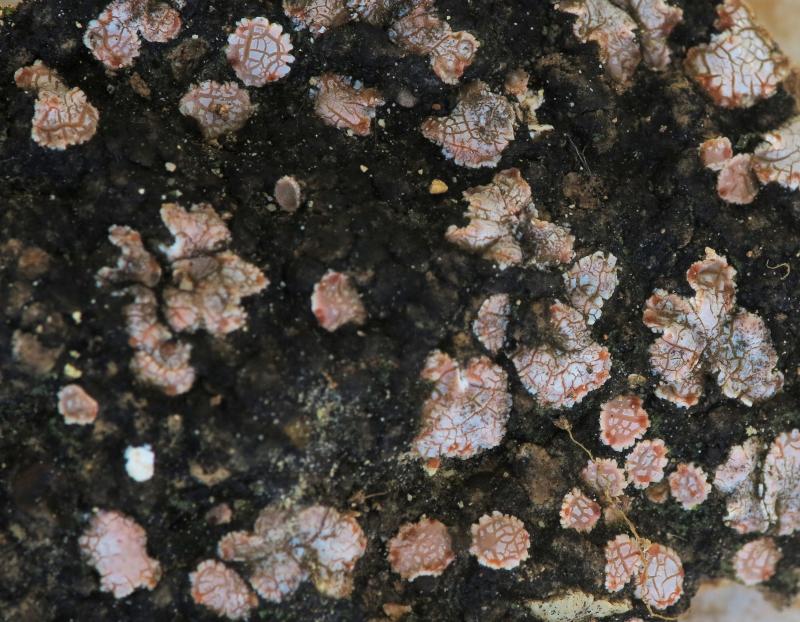
Stephen Martin Mifsud,
31-03-2015 11:03
Re : Pink lichen-like asco
longitudinal cross section revealed 3 layers:
Uppermost (ecternal): pinkish-beige (becomes beige when dry)
Middle layer: green algal layer, thin, well defined
Lower layer: cotton white
Substratum (chalky limestone soil, yellowish) firmly attached below.
Despite a very thin cross-section, I could not see much detail because the section are not squashable and remains several layers thick-compact. Algal cells liberated bur I recall they are not diagnostic for determination. I am afraid I took a very young specimen and not reproductive. I shall now try another section on a mature specimen treated in 10% KOH before Congo red, hoping it will have mature asci.
Interesting adventure!
Tommy Knutsson,
31-03-2015 12:13

Re : Pink lichen-like asco
Psora decipies is a common lichen on that substrate and fits your specimen very well. Young thalli often show that pattern.
Stephen Martin Mifsud,
31-03-2015 12:47
Re : Pink lichen-like asco
Psora decipiens, thank you!
The micro wasn't interesting. Algal cells and many amorphous cells, some branching hypha, and in one small part, tube-like structures with narrow core. Well, good that the macro was enough for determination. Interestingly, unlike ascomycots, there was no micro images of this lichen with a simple google image search.
I will keep the sample & substrate to see how it develops.