28-04-2026 20:07
 Lothar Krieglsteiner
... on twig in the air at standing Ceratonia siliq
04-05-2026 18:13
 Stephen Martin Mifsud
ID request for what seems to be a true aquatic fun
04-05-2026 16:39
Stephen Martin Mifsud
ID request: This specimen was collected in Malta o
04-05-2026 09:50
 Castillo Joseba
Me mandan el material seco de Galicia,(España) re
02-05-2026 12:42
Alain BRISSARDBonjour à tousJeuidi 30 avril dernier on m'a remi
02-05-2026 13:06
Pauline. PennaBonjour Please can someone help me with this id
01-05-2026 22:45
 Thierry Blondelle
Bonjour à tous, Une récolte sur bouse séchée d
14-04-2026 05:32
Ethan CrensonHi all, A few weeks back a friend pointed out som
28-04-2026 20:33
Vitus SchäfftleinHello, I found Trochila ilicina on Ilex aquifoliu
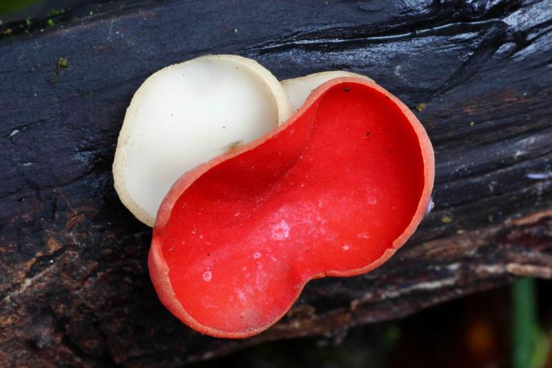
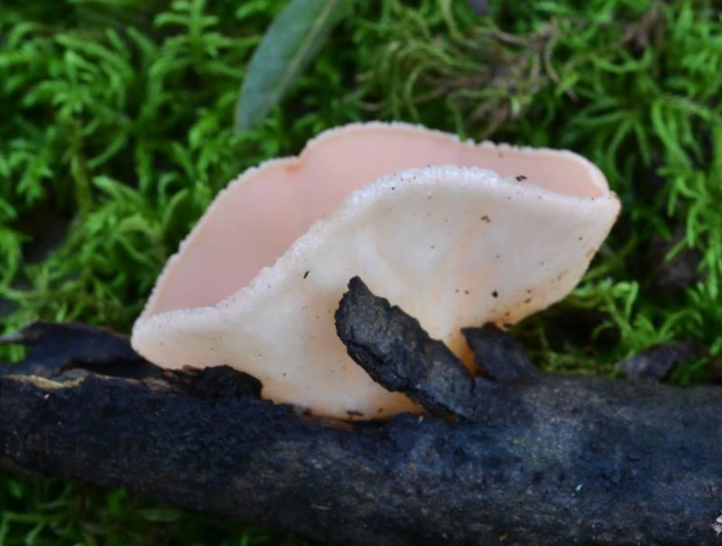
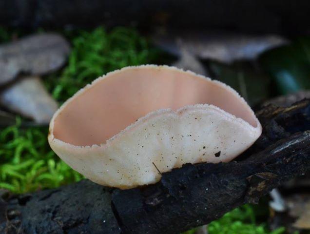
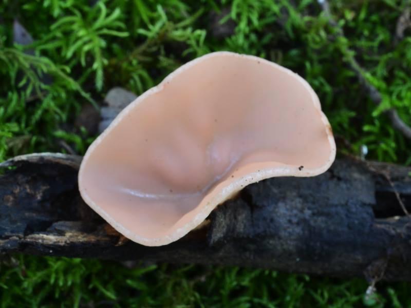
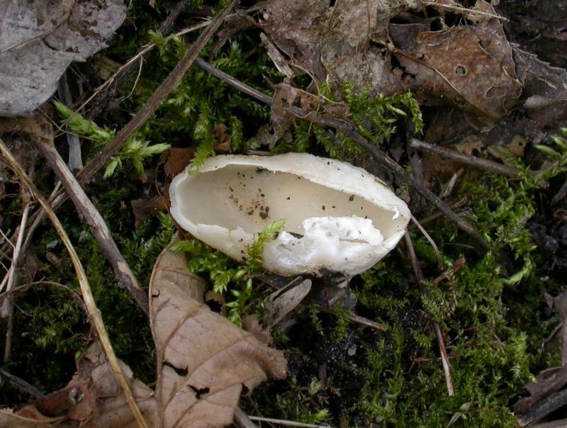
 Dear Friends
I want to show a nice discovery of pink Sarcoscypha coccinea last 1th February on Q. ilex twig, max diameter 3,7 cm. I know orange, orange-yellow, yellow, white coccinea... Someone has already seen the species in this habitus ?
Thank you

I only know Sarcoscypha jurana white.
Good luck.

NL, Flevoland, Eastern part, Reve Abbert, 20.III.05, Fraxinus, in draining ditch.
in the famous Japanese photo-book by R. Imazeki, Y. Otani and T. Hongo, you'll find on p. 555 a picture of a pale pink Sarcoscypha vassiljevae Raitv. which is strikingly similiar to your photograph. I have an article by Raitviir (Eesti NSV teaduste akad. Toimetised, 14,1965: 539-535) describing the species, but it's in Russian, so I can't even find out the colour of the hymenium. In Zotto's master thesis, however, the taxon is mentioned as "pure white" .
According to the Raitviir article, the spores of vassilijevae - 21-26(-29) x 10-13um - are remarkably smaller than those of his S. coccinea (at that time including, probably, austriaca and/or jurana) with 30-40 x 12-15um. Furthermore, the vassiljevae-spore contains one large central oil drop.
The type of S. vassilijevae comes from Wladiwostok ... a long way from your collection site, I'd guess...
Best regards
Till
In my "master thesis" I made a chromatogramm of the red pigment, showing that it consists of a number (three?) of differently coloured substances. I assume that a genetic defect provokes one or two of these being not formed, or even none at all.
In austriaca and jurana I know more of these aberrant collections, partly in mixture with the red ones. But such rose colour I cannot recall.
Yes, vassiljevae i remember white, and I think I translated the Russian text at that time.
Carlo, if you have a microphoto with scale or spore size we could see that your identification is right. On Qu. ilex I only expect S. coccinea with such size.
Zotto

just for curiosity, here is a collection from Japan (presumably by Kiyoshi Iguchi) called S. vassiljevae: http://simocybe.sakura.ne.jp/yosooi-chawan-take.htm
Here some informations I learned with Google translator: apothecia are 3-6cm in dm, "pale yellowish white to pale cream to the entire but, often faintly beautifully tinged with pink", asci 273.8-336.4 × 12.6-13.9 µm, IKI-, spores "surface in oval smooth, colorless in thin wall to moderately thick-walled, and does not react to Meltzer solution, contains 1-2 pieces of oil droplets inside, size 17.1-26.0 × 10.-13.4 µm".
Found on Hokaido and Honshu on partly buried dead branches of Taxus baccata.
Regards
Martin
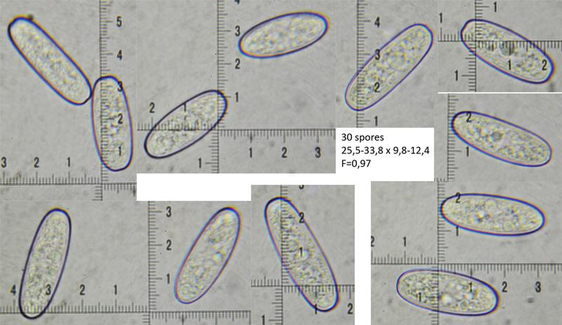
Yes Zotto, spores seems typical to S. coccinea... (25,5-33,8 x 9,8-12,4)
Pink sample grew close many typical S. coccinea samples.
The "genetic defect" concerns already mycelium (?) because the discolored specimens do not never come on the same branches of the typical specimens: they are always isolated.
Clearly the pink specimen must have a low concentration of total carotenoids...Do you think that the higher carotenoid fraction could be the third fraction in your work ? 2' dehydro-plectaniaxanthine-1'ester ?
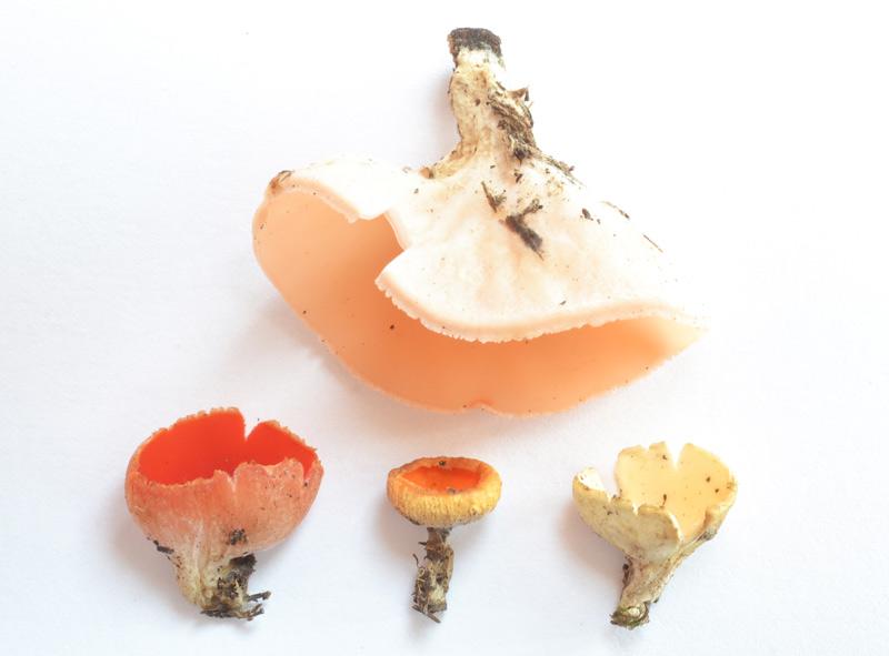
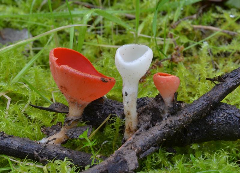
I show you last days collections sent me by a Greek friend with red, orange and white-cream maybe similar to Berthet collection in Arpin ... I add also a complete white 2014 collection (mine).
Best
I did not remember about the chemistry, only that differently coloured carotenoids are involved.
It is too long ago, and I simply compared with the literature. But you may be right with this fraction II. Regrettably I did not photograph the chromatography.
I did not remember about the chemistry, only that differently coloured carotenoids are involved.
It is too long ago, and I simply compared with the literature. But you may be right with this fraction II. Regrettably I did not photograph the chromatography.