
09-06-2026 18:32
Camille MertensSur morceau de roseau immergé 0,5 - 0,7 mm de dia
08-06-2026 17:00
François BartholomeeusenGood day everyone, On June 5 2026, I collected de
08-06-2026 10:16
 Spooren Marco
I don`t have a clou about this fungus,it is not in
07-06-2026 15:10
 William Slosse
Hello everyone,On 05-06-26, I found following asco
05-06-2026 11:02
Thomas Læssøehttps://svampe.databasen.org/observations/10596691
07-06-2026 12:09
 François Freléchoux
Bonjour, Voici une brève description de ce qui m
07-06-2026 12:43
Steve ClementsBojour. This was a strange find on a stick on my
12-07-2015 00:05
 Nedim Jukic
This one from the same locality as the previous on
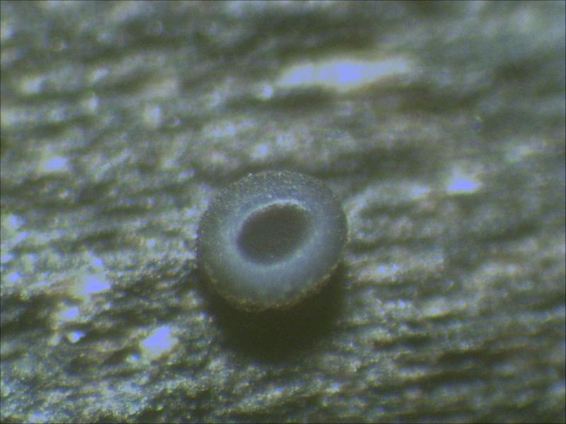
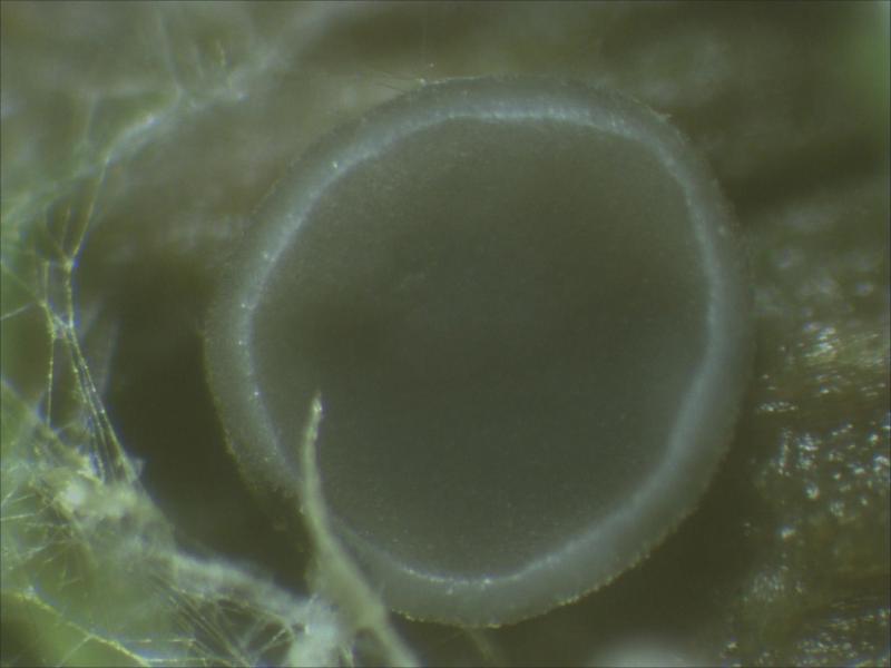
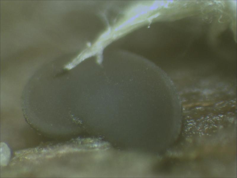
06-06-2026 17:44
Steve ClementsBonjour, This disco was on planed wood 3 x 1.5 cm

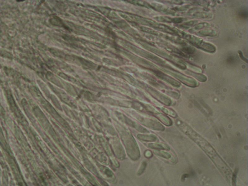
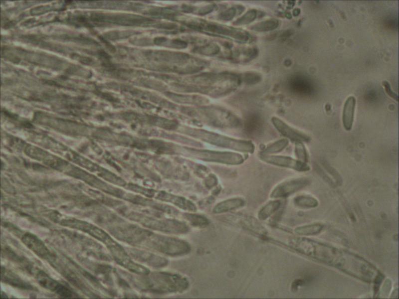
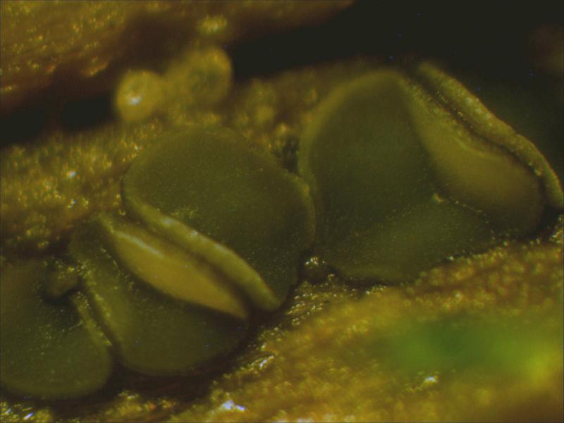
Found on a little peace of wood.
Disk: grey/blue layer with cream coloured edges; when top layer is removed the inside is white.
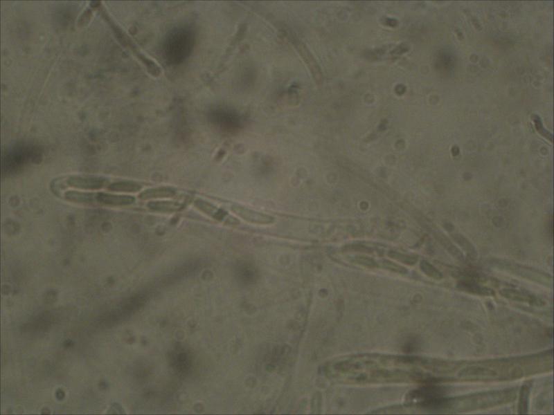
Asco: 8-spored; biseriate in rows of two; 54-62x4-5 um.
Spores: elliptical; 7.5-10.5x2.0-3.0 um
Paraphyses have a slightly rounded top.
The last photo is a little too much on the green side

Hello,
this is a Mollisia species.
No chance to say anything more, many details are lacking.
best regards,
Andreas
Hello Joop,
when I looked at the posting, the last four pictures weren't there ....
But nevertheless, at least the KOH reaction is important, though I believe that it should be negative here. In that case you will end up with Mollisia cinerea s.l., which still is an aggregation of very similar species were I have not the total clue for separating the different taxa.
You can do the KOH reaction macroscopically by putting a fragment of the hymenium in a drop of KOH 20% on a slide which is lying on a white sheet of paper. You then can see a yellow sap yielding from the fragment (KOH positive) - or no change of colour (KOH negative).
You can also do that microscopically by preparing a small piece of the hymenoum in a water mount and then adding KOH at the side of the cover slip. You can watch through the microscope when the KOH reached your piece of hymenium: The vacuolar bodies in the paraphyses will be dissolved rapidly by the KOH reaching these paraphyses. The dissolving will either result in a yellow colouration (positive) or the mount will stay completely colourless (negative).
best regards,
Andreas