

17-05-2026 19:05
Thomas FlammerI have found this tiny 200 ym cup shaped apothecia
17-05-2026 16:41
Margot en Geert VullingsWe found this Lachnum on an old Rubus stem.Fruitbo
05-04-2026 22:46
 Lothar Krieglsteiner
on wood of Ceratonia, Algarve, 3.4.2026.The color
15-05-2026 13:33
Sylvie Le GoffBonjour à tousJe serais très reconnaissante enve
16-03-2011 14:31
roman vargas albertoHi. I would like some opinion about this Peziza
14-05-2026 05:36
Ethan CrensonHi all, I haven't paid much attention to Lachnu
10-05-2026 23:17
 Andreas Gminder
Hello,today we found in a moist steep decidous for
11-05-2026 12:32
 Bernard CLESSE
Pourriez-vous m'aider à identifier cette héloti
13-05-2026 15:26
 François Freléchoux
Bonjour,Voici une récolte faite il y a quelques j
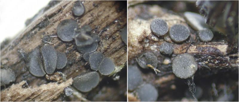
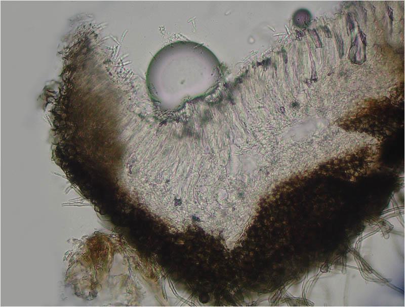
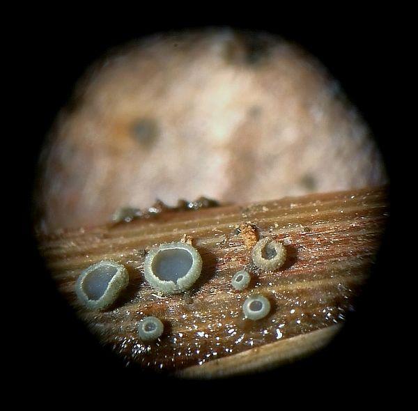
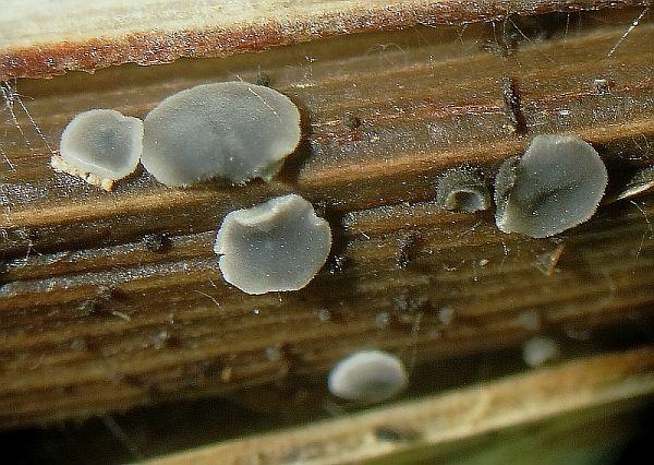
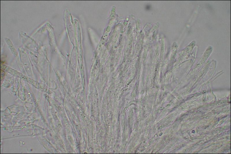
I collected these apothecia of grayish color in the hymenium and dark brown in excipulo.
The reaction with KOH is negative.
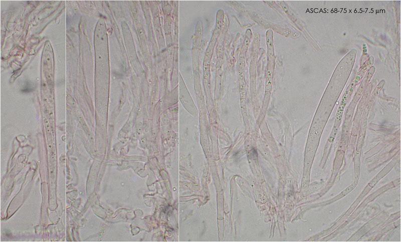
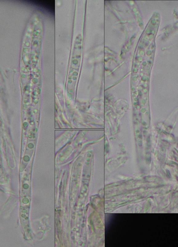
Asci are IKI + (blue). 68-75x6.5-7.5um size. Croziers +
Spores with a septum (difficult to distinguish in water, but visible in IKI).
Given the features mentioned, I think it may be mollisia humidicola. Can anyone confirm or refute the determination?
Thanks in advance, greetings
Susana
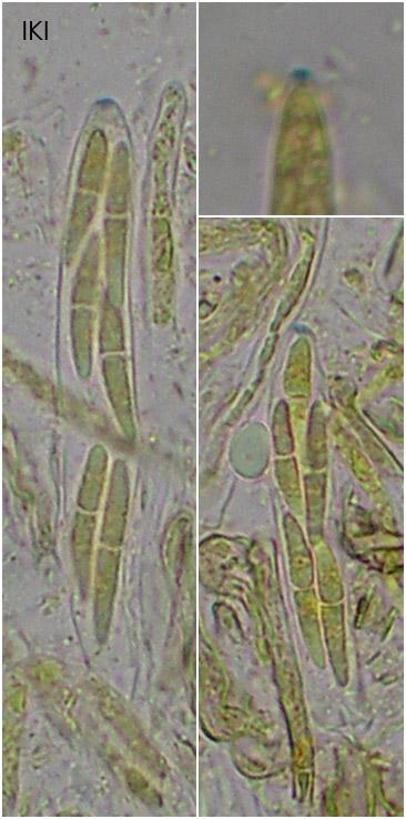
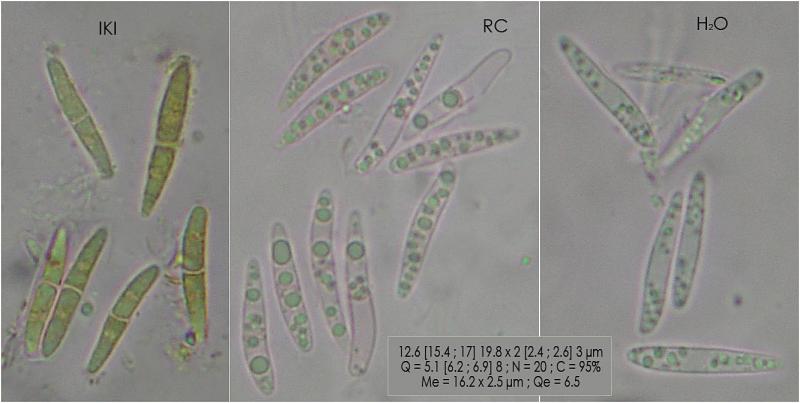
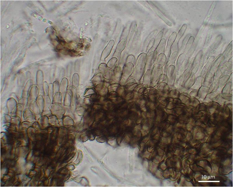

You have also nonseptate spores on your photos, those in which an "empty" space is in the middle (the nuclear region). When the oil drops are found in the centre then there is also a septum.
Tell me please what was the substrate, indet. grass?

Mollisia humidicola/ luctuosa is in our distrikt a frequently species on grasses. This species has been found on
Calamagrostis epigejos, Carex spec., Carex rostrata, Juncus effusus, Molinia coerulea, Phalaris arundinacea, Scirpus sylvaticus.
Greetings
Peter Püwert.
thanks
I do not know what type of substrate is, but it is not a grass. It is a piece of hardwood branch, collected in an area where they grow elms.
Saludos
Susana
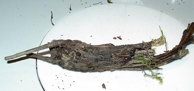
could you please try to make a cross cut of the substrate where it is not too rotten, i.e., where a section is still possible? It must not be a microscopic section, but only a surface view on the wood. Wood is rather easily distinguished from herbs/monocots.
It would be a great surprize to see a species with siuch long, septate spores on ligenous substrate. The diameter of the piece seems to lie at about 15-20 mm, isn't it? There is no large grass with such thick base?
Zotto

Regards
Martin
Saludos
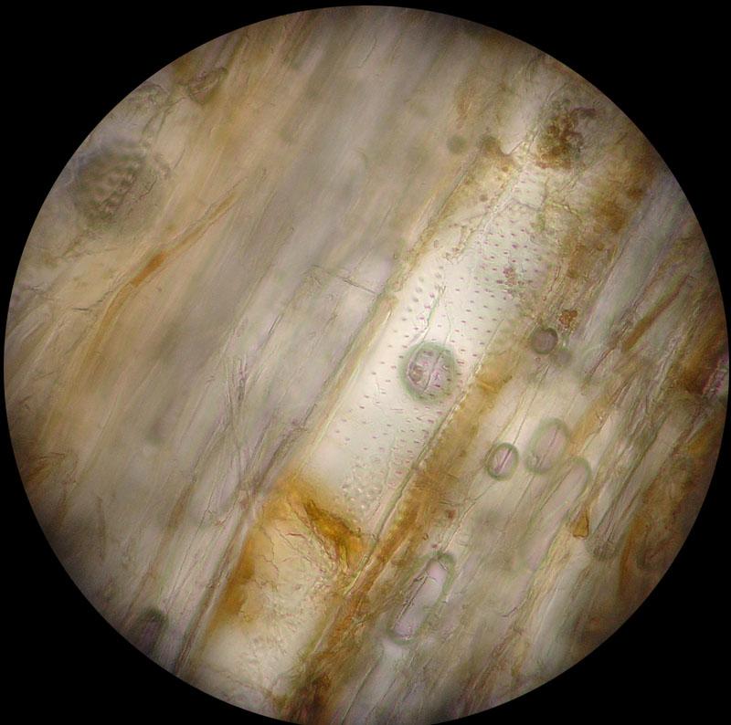
This is a cross section of the substrate.
Un saludo
Susana
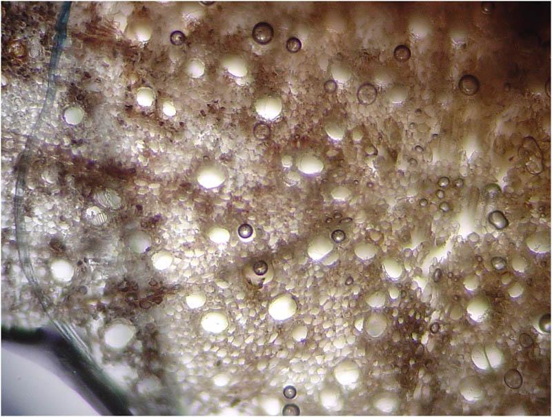
I assume the substrate is rather hard, is it? This section covers only about 2 mm of the centre, does it? How thick is the entire stem?
Ulmus seems completely excluded. Salix could be a possibility, but I would expect a parenchymatic inner region that I do not see here.
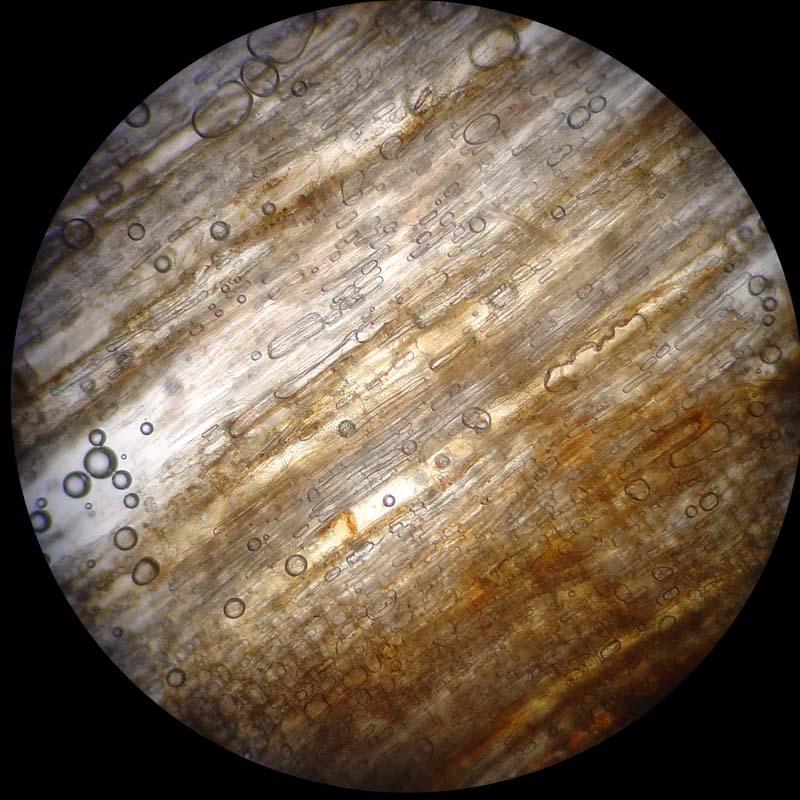
I do not want to bother you, but now that you got such a fine cross section, a radial section would be useful to see the pore perforations and the pits.
Zotto
Yes, the substrate is hard.
Yes, the section is about 2mm
Entire stem is 15mm, but it is very deteriored.
I send you a pictures from longitudinal cut. I hope the pictures will help you. I do not know what needs to look
Susana
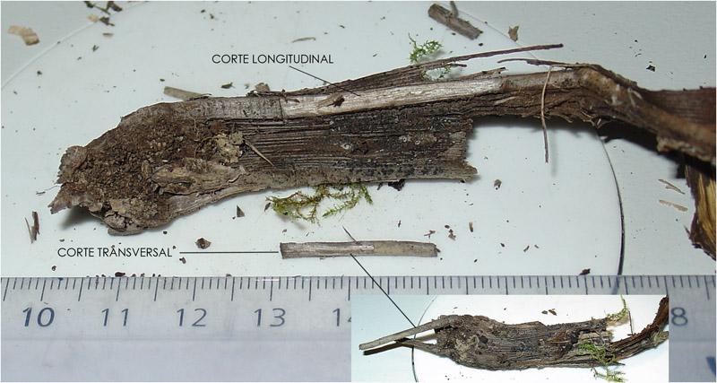
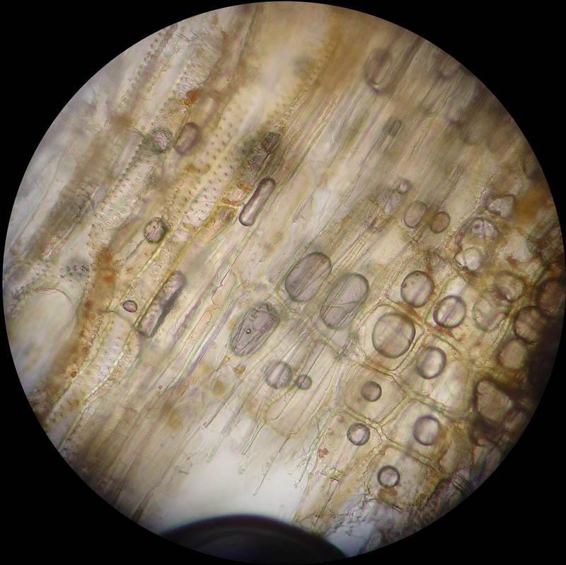
Helpful would be to have a section through the 15 mm thick part. I assume this is difficult, but it is enough to know whether the interior is hollow, and how thick the wall. The surface looks rilled, does it?
You're right. Effectively, the interior is hollow and the surface is rilled. The wall thickness is about 3.5mm. I send you a photo of the cut.
a greeting
Susana
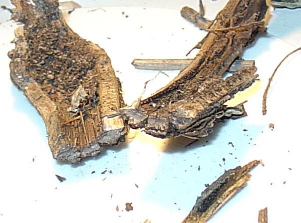
That would fit to the substrate!
Zotto
Susana
Within dead asci the spores often remain alive and finally develop a septum or become brown etc.
You did not make a water preparation of the fresh fungus without applying much pressure? Then we could also see the living paraphyses.
Effectively, the paraphyses are not mollisia type.
I see septate spores inside the ascus.
What do you think?
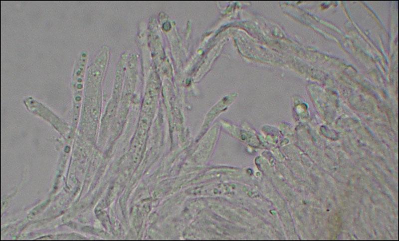
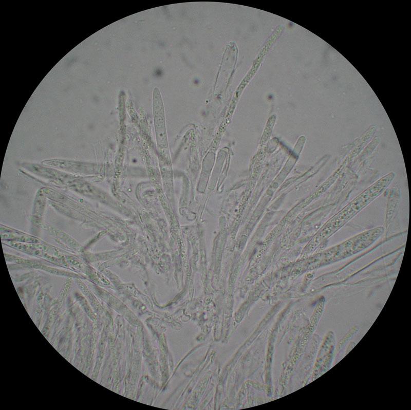
The asci are completely dead. Living asci look very different , much wider. If you had dried the sample then no wonder when you find mainly dead elements. Asci are most sensitive to drying.
Saludos
Susana