

05-05-2026 22:40
Gernot FriebesHi,I believe this is a Plagiostoma growing on a Sa
06-05-2026 11:25
 Castillo Joseba
Me mandan el material seco de Galicia (España) re
06-05-2026 17:23
Thomas Læssøehttps://svampe.databasen.org/observations/10594257
28-04-2026 20:07
 Lothar Krieglsteiner
... on twig in the air at standing Ceratonia siliq
04-05-2026 18:13
 Stephen Martin Mifsud
ID request for what seems to be a true aquatic fun
04-05-2026 16:39
Stephen Martin Mifsud
ID request: This specimen was collected in Malta o
28-07-2011 18:31
 Alex Akulov
Dear FriendsToday I made the pdf file of Velenovsk
04-05-2026 09:50
Castillo Joseba
Me mandan el material seco de Galicia,(España) re
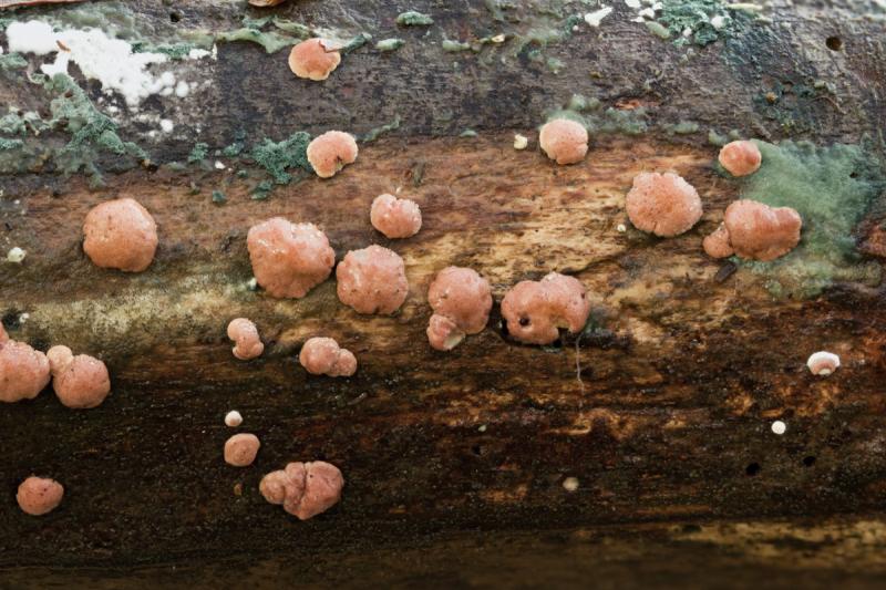
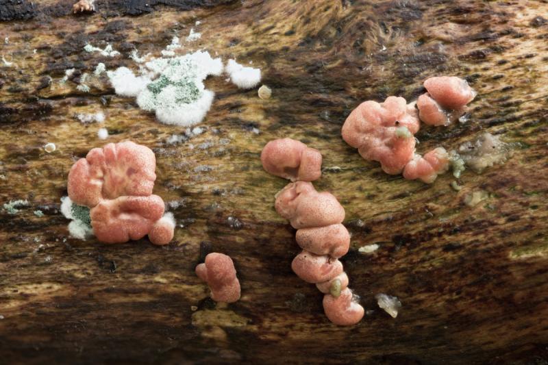
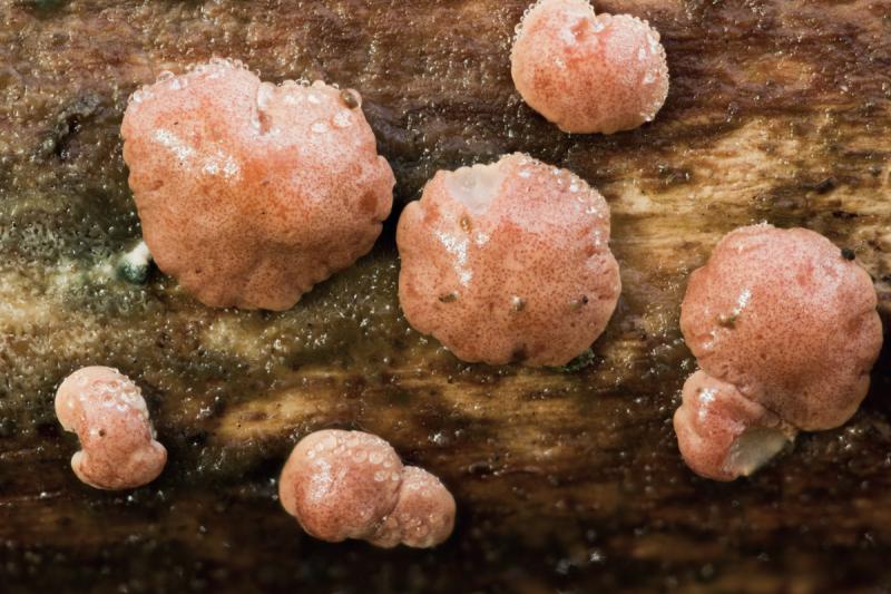
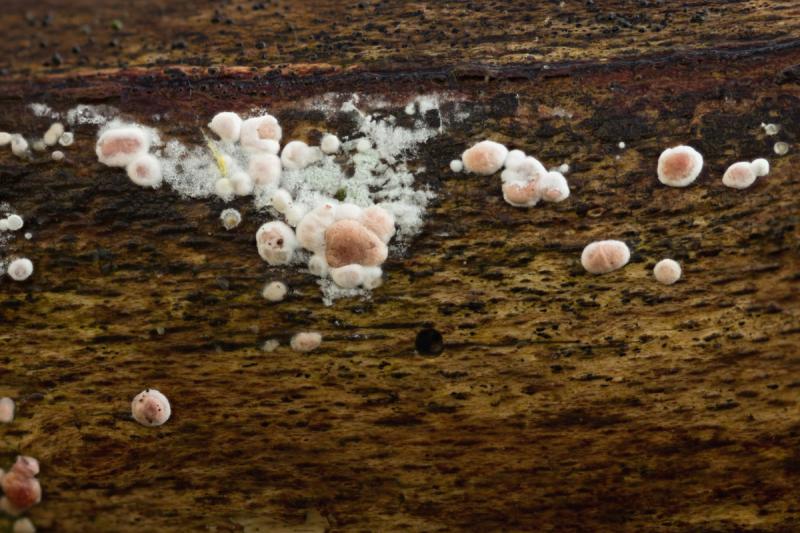
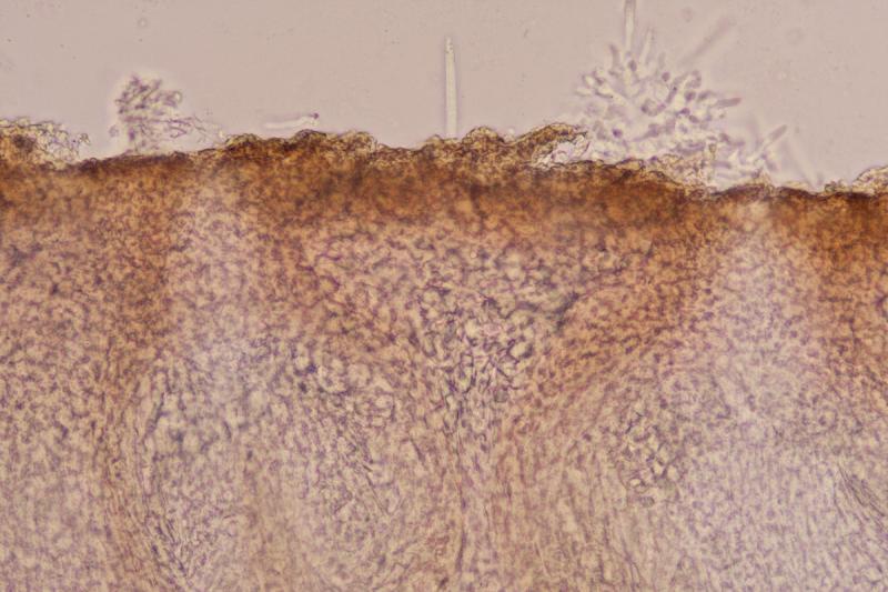
Here is what I believe is a Hypocrea found on Fagus branch on the forest floor. The stromata are not mature but the green anamorph was present which may help to identify it. The stromata were to 5mm and there were many of them. Attached to the substrate right up to the edge. They were a pinkish colour with visible darker ostiolar openings creating a pattern of dots on the surface. They dried to a pale brown.
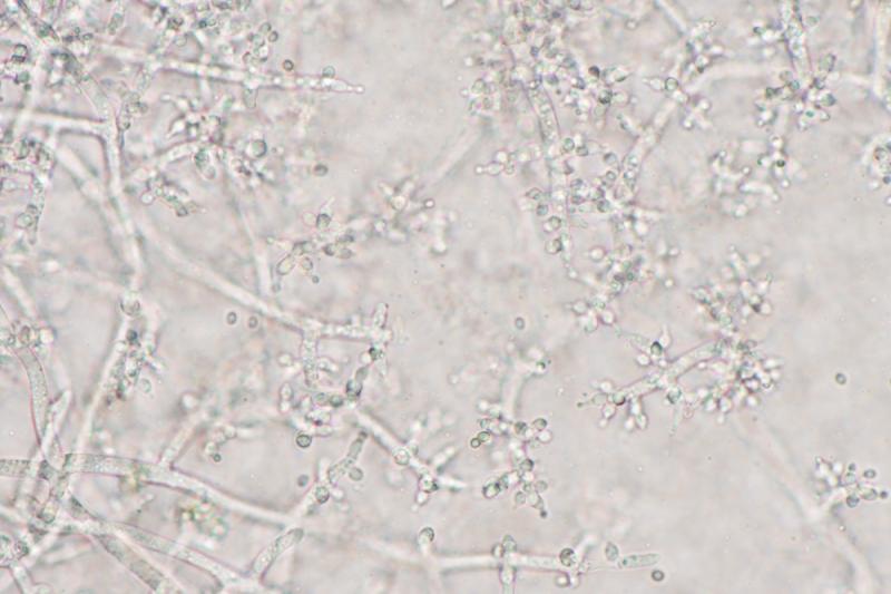
The anamorph was green and I don't have the language to describe the structure of the conidiophores and phialides, hopefully the pictures will help
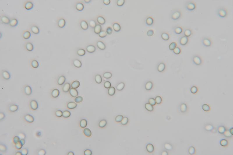
The conidia were smooth and ellipsoid 3.3 X 2.4 QE 1.4
My attempts to key it give Hypocrea minutispora and that does look OK with the structure of the conidiophores and phialides as described for that species in this paper although the conidia may be a bit on the large size.
If anyone can help I would appreciate it.
Many thanks
David
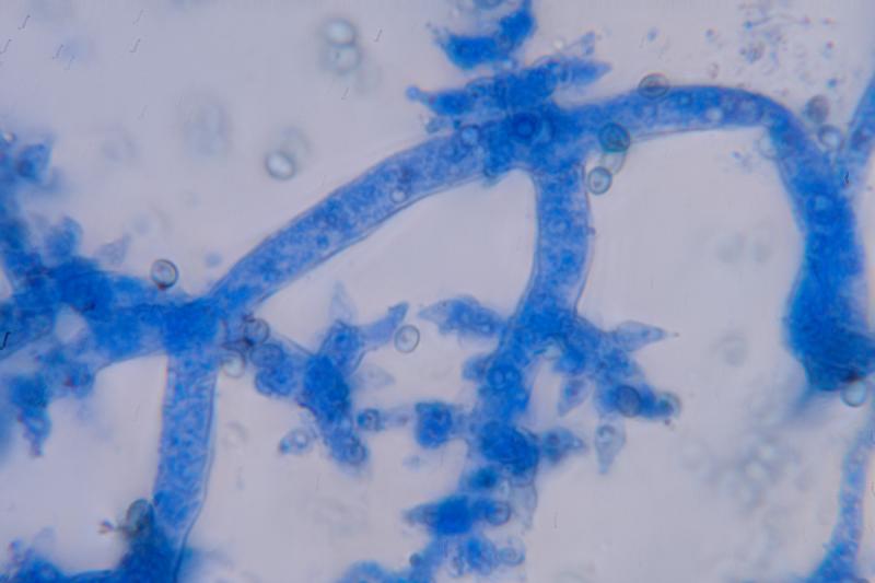

I think your fungus is H. minutispora but it would be necessary to compare it to H. pachybasidioides.
Christian
Thank you Christian,
I looked again at the paper I mentioned in my first post and it appears that H. pachybasidioides is in the "polysporum" clade and its anamorph, (Trichoderma Polysporum) has white/hyaline conidia where my specimen had green conidia. It looks like it also has infertile, corkscrew-like extensions of the conidiophores unlike my specimen. The green, smooth, ellipsoid conidia, along with the structure of the conidiophores pictured in the paper above, do seem to leave only T. minutisporum / H. minutispora.
Regards
David