19-05-2026 10:27
Patrice TANCHAUDBonjour, récolte récente sur terre retournée i
04-06-2026 10:50
 François Freléchoux
Bonjour, J'ai trouvé hier un petit asco observé
04-06-2026 07:02
François Freléchoux
Bonjour, Voici la description d'une espèce qui p
04-06-2026 13:34
Gernot FriebesHi,I am interested to hear your opinion on this Le
04-06-2026 11:36
Gernot FriebesHi,found on Vaccinium myrtillus.Asci: IKI –, 8-s
22-05-2026 13:29
Gernot FriebesHi,I am curious to hear your opinion on this mater
18-10-2022 00:12
Valencia Lopez Francisco JavierHola amigos/asRecientemente encontré esta colecci
 Hi everyone,
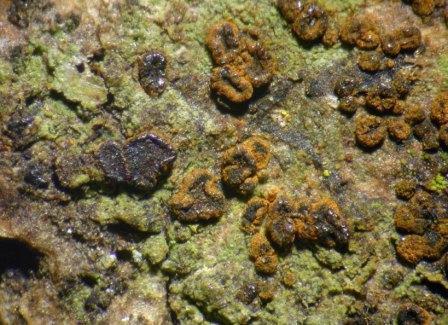
I got a fungus on bark from southern Italy, seemingly with no connection to the surrounding lichens. If it were lichenicolous, I would presume it to be a Buelliella. Has anybody any idea what this could be?
Ascomata sessile, constricted below, initially closed, later apothecioid, up to 0.5 mm diam., irregularly roundish, disc black but covered with a rusty pruina, margin prominent, densely rusty pruinose.
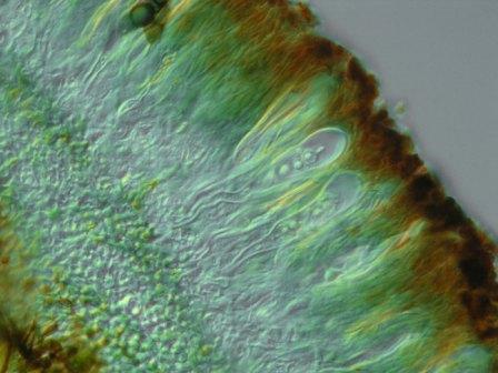
Hypothecium brownish, 55 µm high, hymenium hyaline below, brownish above, 120 µm high, epithecium brown, covered with dark brown granules, excipulum dark brown, up to 50 µm thick.
Paraphyses septate, sparsely ramified, 2–2.5 µm wide, hyaline below, brownish above, the upper cell sometimes enlarged up to 4 µm, brown in the upper half.
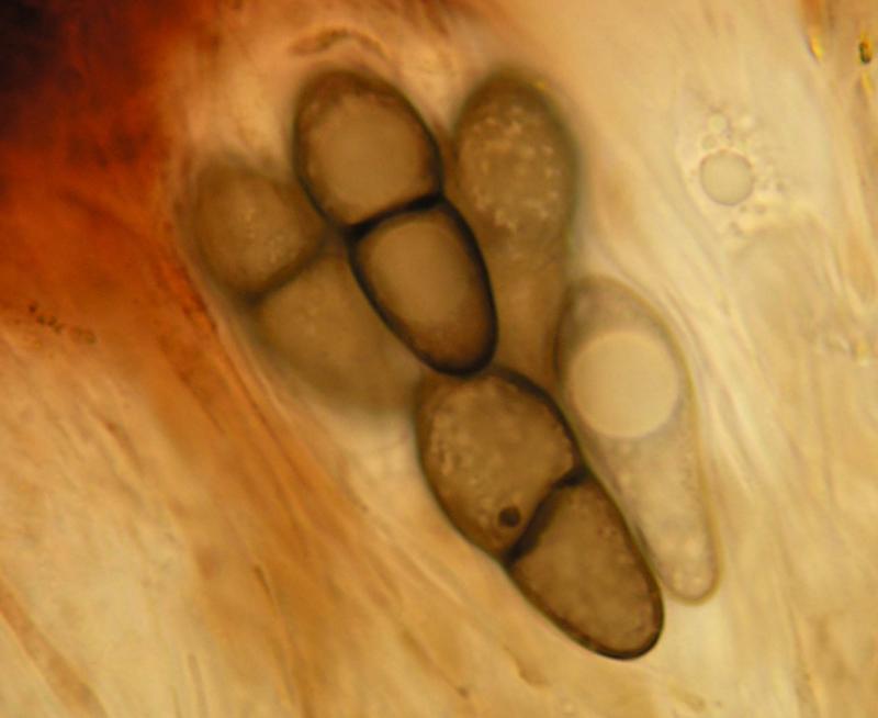
Asci clavate, 80–95 × 15–26 µm, apically thickened, with an internal beak, with a long stalk, 4–8-spored.
Ascospores 1-septate, grey, the upper cell rounded or slightly attenuated, the lower attenuated, narrower than the upper one, constricted at the septum, with one big guttule in each cell, surface ± smooth but appearing foveate, (20–)20.8–23.1(–24) × (8.5–)9–9.9(–10) µm, l/b = (2–)2.2–2.5(–2.6) (n = 20).
Pruina K+ violet, not dissolving. Hymenium above K+ grey, I+ reddish. Asci externally I/KI+ pale blue, no I/KI reacting apical structures.?