
08-06-2026 10:16
 Spooren Marco
I don`t have a clou about this fungus,it is not in
08-06-2026 17:00
François BartholomeeusenGood day everyone, On June 5 2026, I collected de
07-06-2026 15:10
 William Slosse
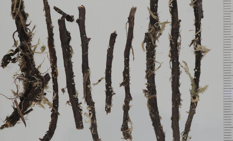
Hello everyone,On 05-06-26, I found following asco
05-06-2026 11:02
Thomas Læssøehttps://svampe.databasen.org/observations/10596691
07-06-2026 12:09
 François Freléchoux
Bonjour, Voici une brève description de ce qui m
07-06-2026 12:43
Steve ClementsBojour. This was a strange find on a stick on my
12-07-2015 00:05
 Nedim Jukic
This one from the same locality as the previous on
06-06-2026 17:44
Steve ClementsBonjour, This disco was on planed wood 3 x 1.5 cm
14-08-2016 23:15
 Alex Akulov
Dear friendsCan you help me to find the descriptio
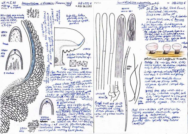
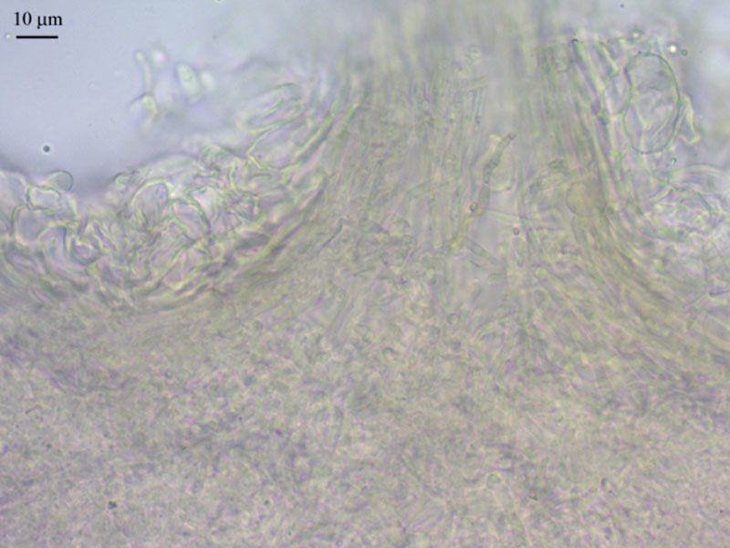
It is picked from sphagnum bog upper layer and at dead branches of Chamaedaphnethere were apothecia in quite abundance (N61,054422° E69,456725°).
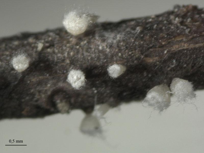
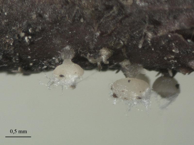
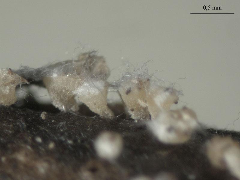
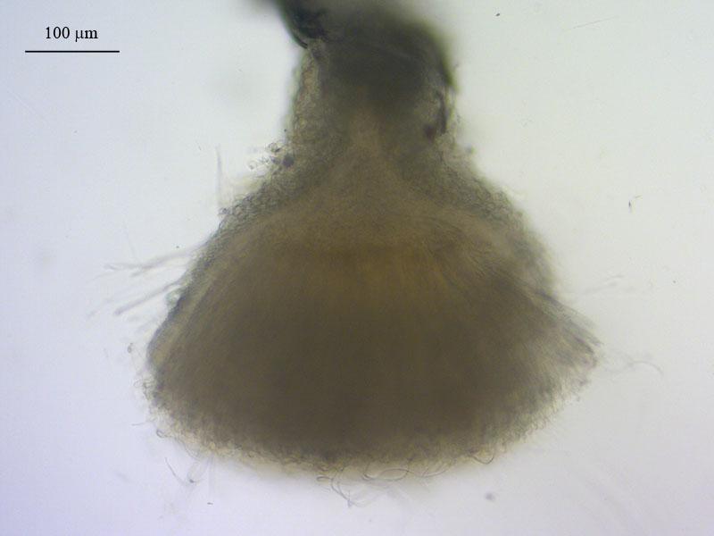
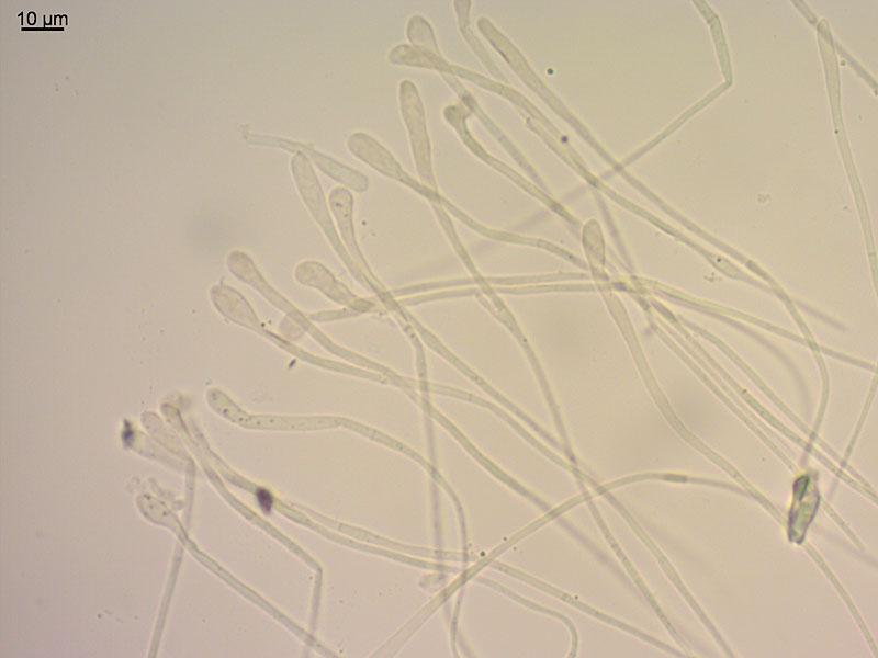
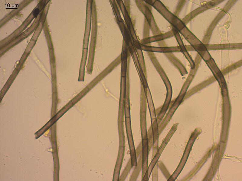
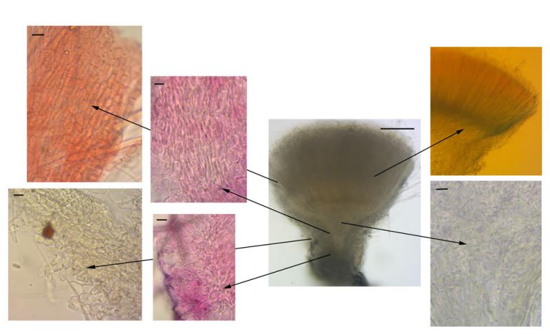
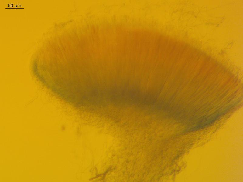
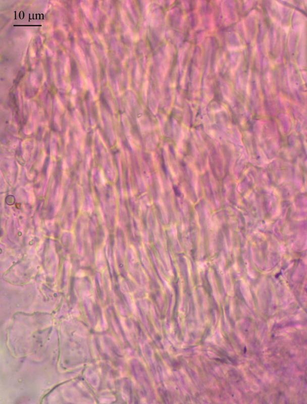
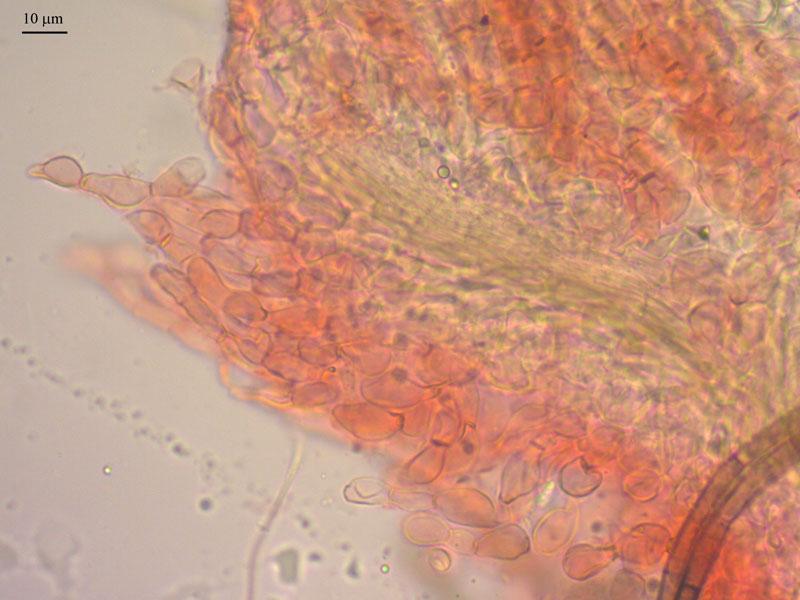
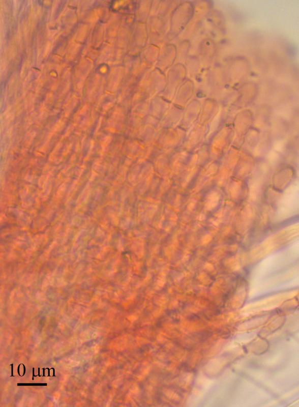
Apothecia sessile or with short thick stipe, up to to 500 mk high, receptacle white to yellowish, disc to 500 mk broad, convex, with pure white cottony layer of spores, seat on conspicuous dark brown subiculum.
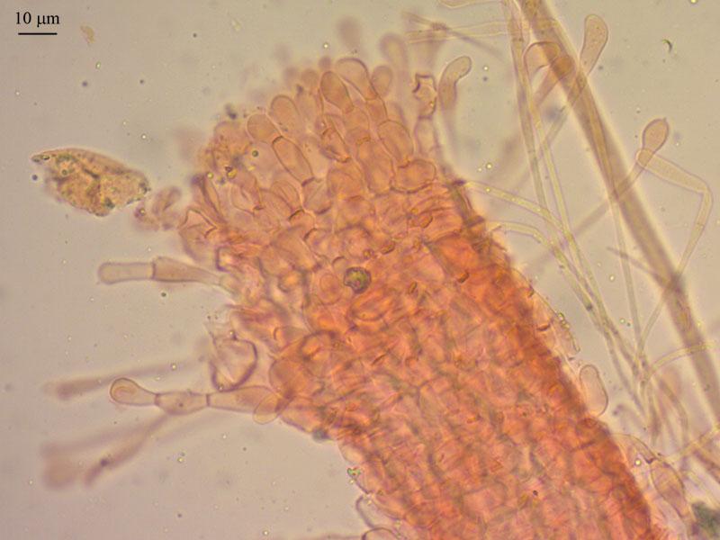
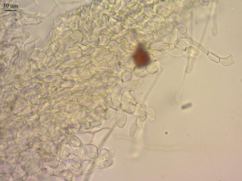
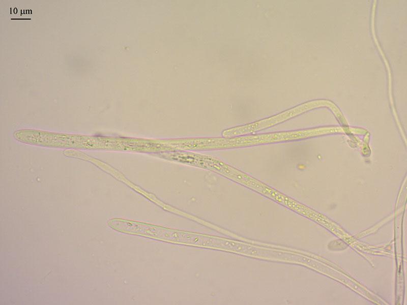
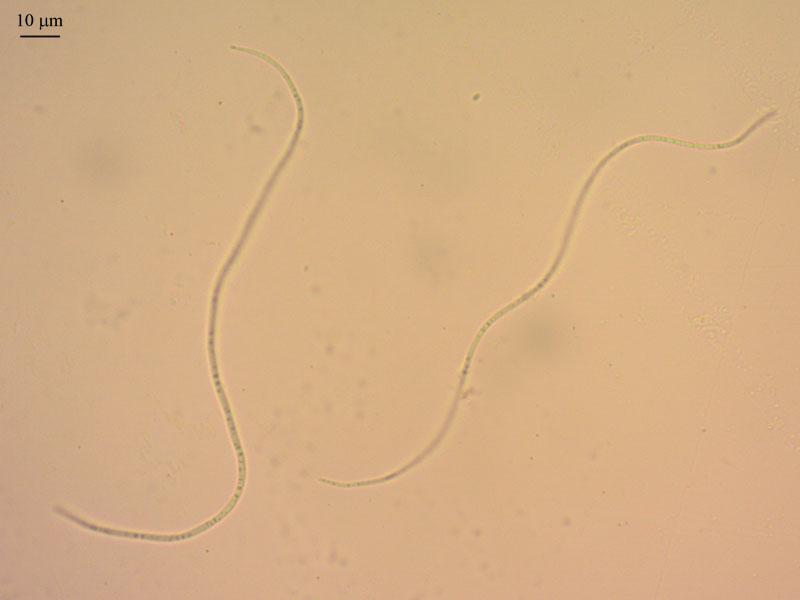
Ectal excipulum from textura globosa (outer layers) to prismatica (inner layers), cells about 15 mk in diam, medullary excipulum from textura intricata, cylindrical septate hyphae about 2 mk broad, outer layer of excipulum and edge with "hais" (chains of 4-5 cells, constricted to moniliform, end cells clavate); subiculum hyphae cylindrical, stright, septated, 4,5 mk thick, brown; asci long and narrow, gradually enlarged from base (2 mk) to obtuse-conical tip, 188 (177-213) x 7,5 (6,5-8), clamped, with small amyloid pore (1 mk); paraphyses cylindrical, segmented, clavate at the end segments, which exceed the asci, branching not checked, near 2 mk broad in middle part, 5,6 (4-11) mk at enlarged tips; spores filiform, with upper end slightly obtuse, base end narrowed (not always seen), segmented (better seen in MLZ, about 15 septa), circinate, with tiny guttules, not disarticulating, not germinating, mean length 184 mk (5 strightened spores measured, but impossible to do presicely measure), 1,6 mk broad.

You can try to see the amyloid reaction at the margin, also I assume yours lack the crystals I have seen on the exterior of the flanks.
Zotto
Yes, there is clear reaction of MLZ in inner layer of excipulum ("perihymenial zone") and no reaction in medullary excipulum.
No crystals, i observed in water and looked at dry apothecia, which may be help, but have not found them (probably, it may depend of wetness of habitat?).
And i tried to improve description of tissues in my specimen, since there are some differences in the drowing in the paper of Beton and Weste (1980), and your drowing, information about excipulum features also provided in Sanches and Korf (1966) for the sections of Vibrissea.
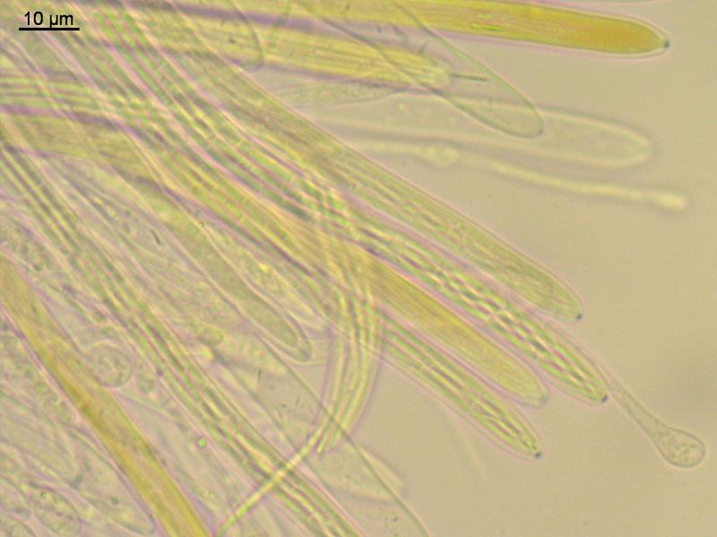
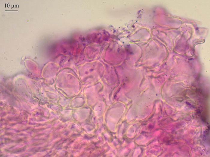
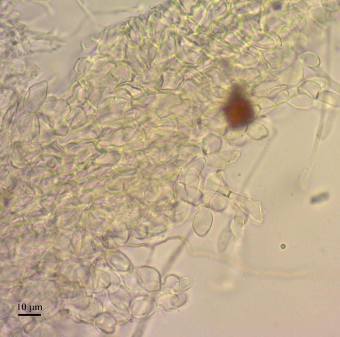
Ectal excipulum from textura angularis-prismatica; at the base cells large, angular, isodiametric, thick-walled, with longitudinal axes perpendicular to the outer surface; in the middle and up to the margin cells in the inner layer becoming elongated, laying parallel to the outer surface and yet thick-walled; outer layer from cells with thinner walls, ellipsoid to prismatic, arranged in rows, with free rejected, hair-like chains from 4-5 cells scattered at all length and densely covering the margin.
Medullary excipulum from textura intricata, hyphae about 2 mk broad; becoming parralel when approaching the excipulum.
I think it worth to draw these tissues when i will be more skillfull in drowings; now prepared some more pictures.
Yet i did not have the last two papers, probably usefull since the key for species is there.
Beaton, G., Weste, G., 1980. Vibrissea: Two North American species redescribed and a new Javan species. Transactions of the British Mycological Society 75, 243–248.
Sanchez, A., Korf, R.P., 1966. The Genus Vibrissea, and the Generic Names Leptosporium, Apostemium, Apostemidium, Gorgoniceps and Ophiogloea. Mycologia 58, 722.
Beaton, G., Weste, G., 1976. Australian discomycetes: Log-inhabiting Vibrissea species. Transactions of the British Mycological Society 67, 129–132.
Beaton, G., Weste, G., 1977. Australian discomycetes: A new Vibrissea species. Transactions of the British Mycological Society 69, 323–325.?
Crystals are usually a good character in the Helotiales, but sometimes they are absent though the species shoul have some.
Here you can download my drawing in better resolution.
https://www.cubby.com/p/_778a6f3587954e008790666f822f8e4e/7a+Helotiales#7a%20Helotiales/Mollisiaceae/Vibrisseae
There are also macros by Jens Petersen (files 122-Leucovibrissea obconica JHP-10-137-711_07282010.jpg):
Leucovibrissea obconicaNorthern Europe, Bjurälvens naturreservat, (64.927888 / 14.144511), vegetation type: meadow, associated organism: grasses, substrate: stems, grasses, etc., leg.: Thomas Læssøe & Jens H. Petersen, det.: Thomas Læssøe & Jens H. Petersen, coll. number: JHP-10.137.Remarks: sp. 165 x 1 µm.
Zotto

You can find the missing papers on the following link:
http://www.cybertruffle.org.uk/cyberliber/index.htm?
Kind regards,
Ivana