

22-04-2026 20:54
 Enrique Rubio
Hi to everybody.This Pyrenopeziza grew in moist le
24-04-2026 03:16
 David Chapados
Found while looking at something else from wood in
22-04-2026 01:06
 Richard VALERI
Bonjour à tous.Je vous présente cette Nectria s.
22-04-2026 20:17
 Marian Jagers
Is anyone familiar with the Hyphomycetes genus Pse

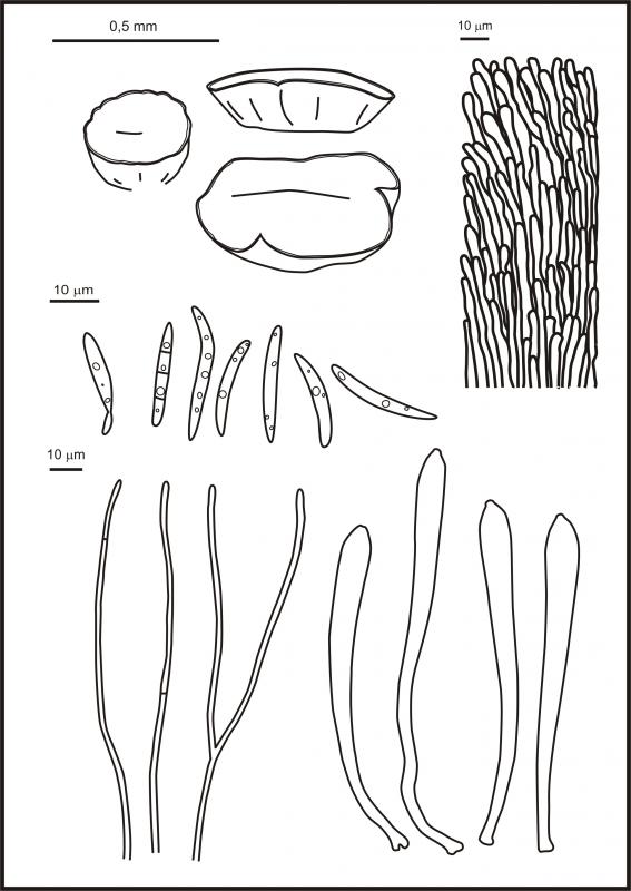
i would like to post some findings from West Siberia, if it is appropriate:
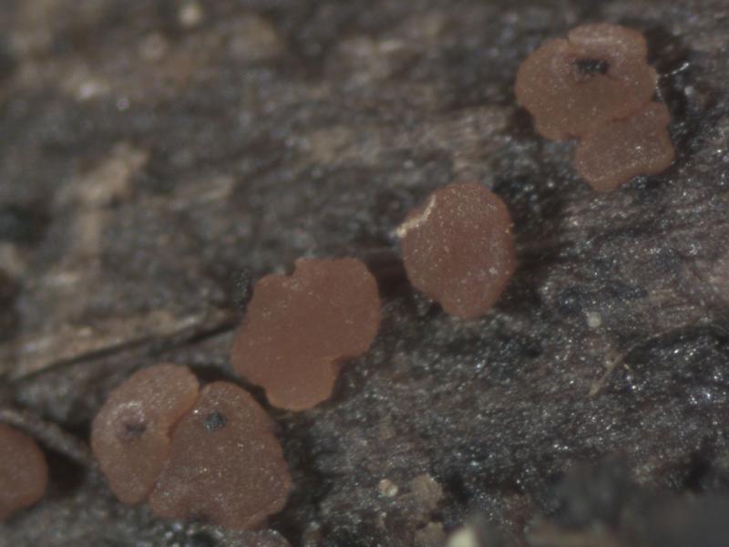
Durella cf. atrocyanea? (but the spores are narrower, probably undeveloped since most were inside the asci).
Was collected on decorticated pine branch (Pinus sylvestris), N61,054422° E69,456725°.
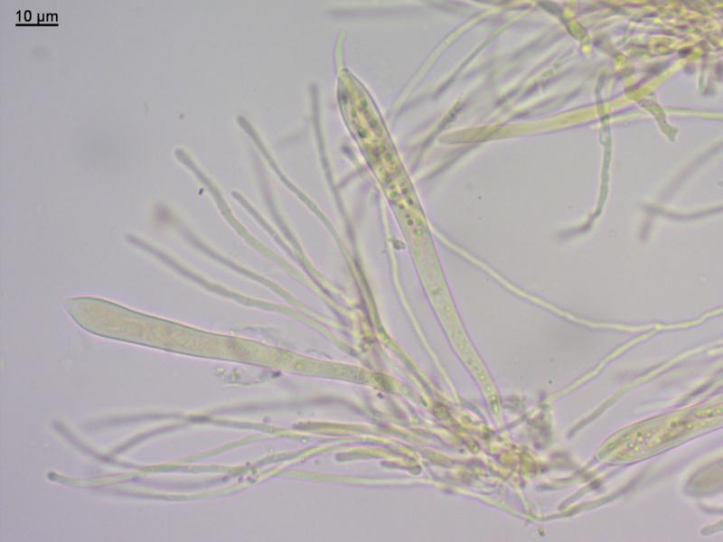
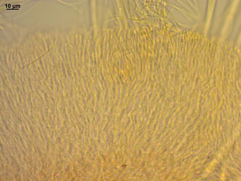
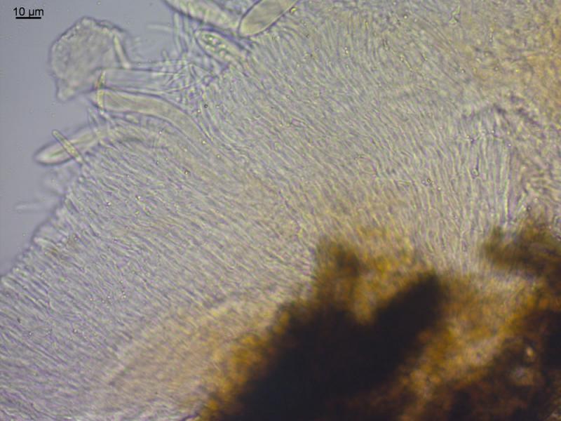
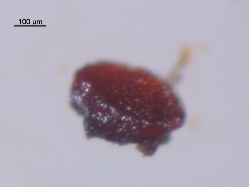
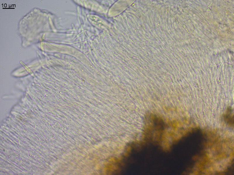
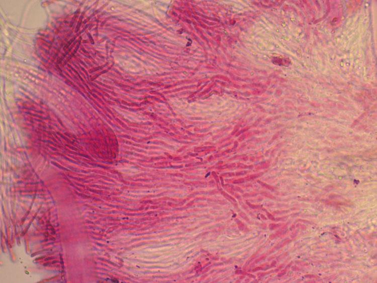
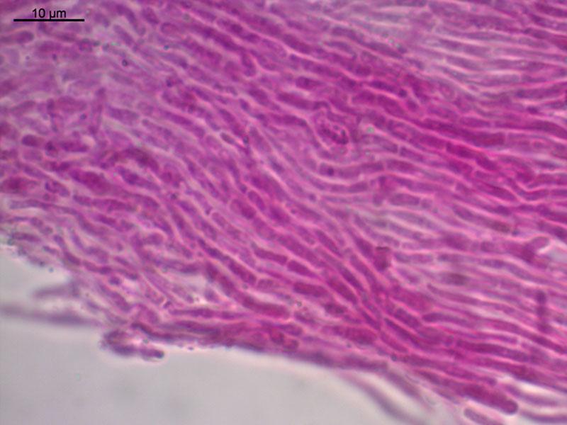
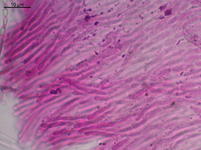
Frb pulvinate-cupulate, reddish-brown, up to 500 x 150 mk, hymenium surface flat or convex, smooth, outer surface smooth, the same color, brownish at the base.
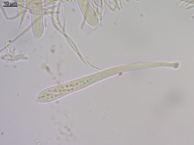
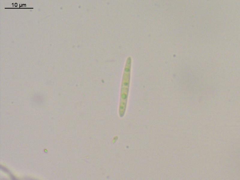
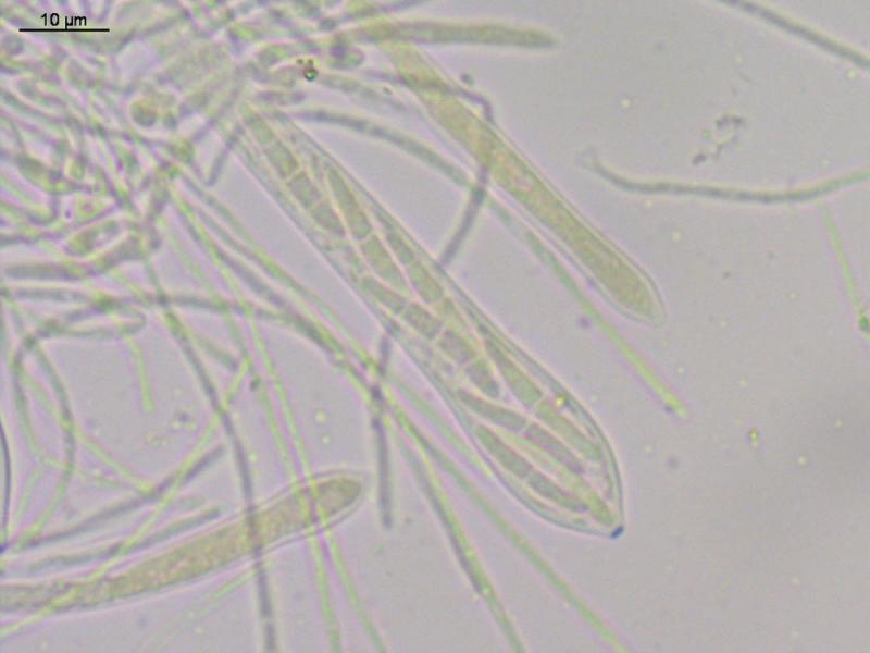
Excipulum from textura porrecta, hyphae at the edge obtuse, not enlarged; asci large, enlarged to the tip, tip conical, with amyloid pore, with scarce clamps, 117 (104-135) x 9 (8,3-10,5) mk; spores fusoid, some bent, some obtuse from one end, 1-3 segmented (well seen in melzer), with several round guttules, 18,7 (14,2-23) x 3 (2,5-3,5) mk [10 spores: mean, min-max]; paraphyses cylindrical, branched, 1,5 mk broad.

D. atrocyanea has rounded inamyloid ascus apices. Yours look as having a small blue apical ring?
Zotto
excipulum from cylindrical thick-walled hyphae (brownish), tightly arranged, and with short free ends at the edge (not hairs).
I have Gorgoniceps aridula in my collection, compared asci - yes, they look quite similar. Though spores really longer, 60-95 mk in G. aridula, - (i will check other species, may be there something will match).
Zotto
Also found that frbs are very bright red in transmitted light, and red pigment saturated even in water in the mount.
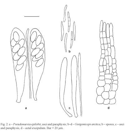
There is link to this species in the Raitviir's paper (2008, the Helotiales of the Magadan and Chukotka areas of the Russian Arctic):
"This short spored species of Gorgoniceps [G. arctica sp. nov.] differs from G. hypothallosa, which has spores of the same size, in much paler apothecia, which do not turn red when mounted in KOH. Two other lignicolous species of the genus, G. aridula and G. viridula, have much longer spores and like G. hypothallosa grow on coniferous wood."
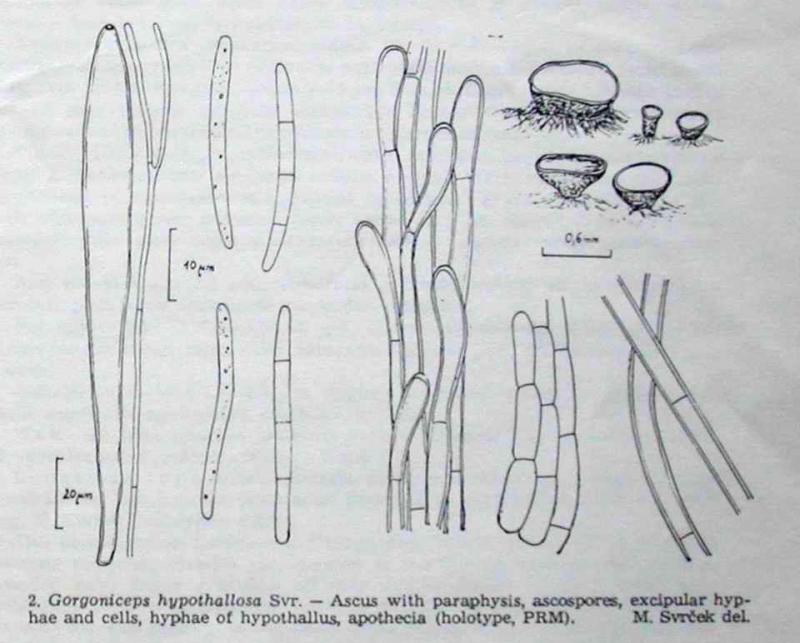
Unfortunatelly, the description of G. hypothallosa not available for me right now (Svrcek M.: New or less known Discomycetes. XIII. Ceská Mykol. 38(4): 197 (1984).
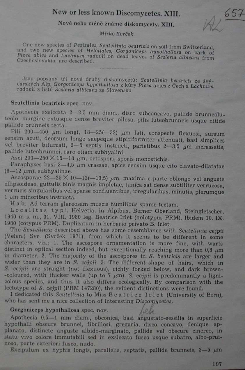 Svrcek-1984-New-or-less-13-1-0001.JPG
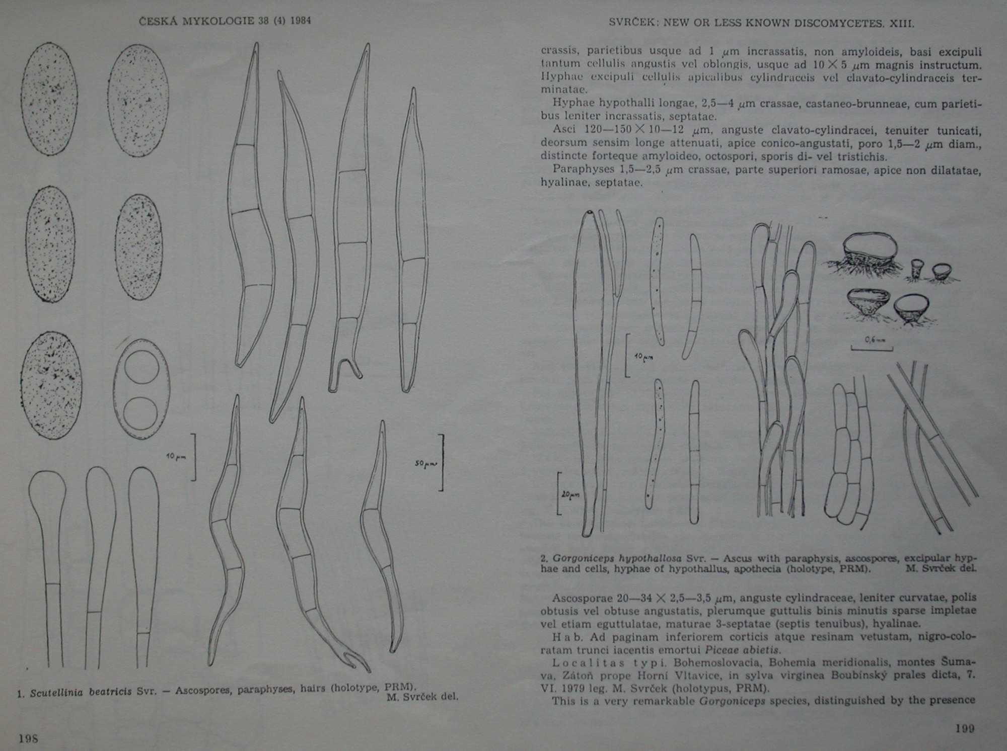 Svrcek-1984-New-or-less-13-2-0001.JPG
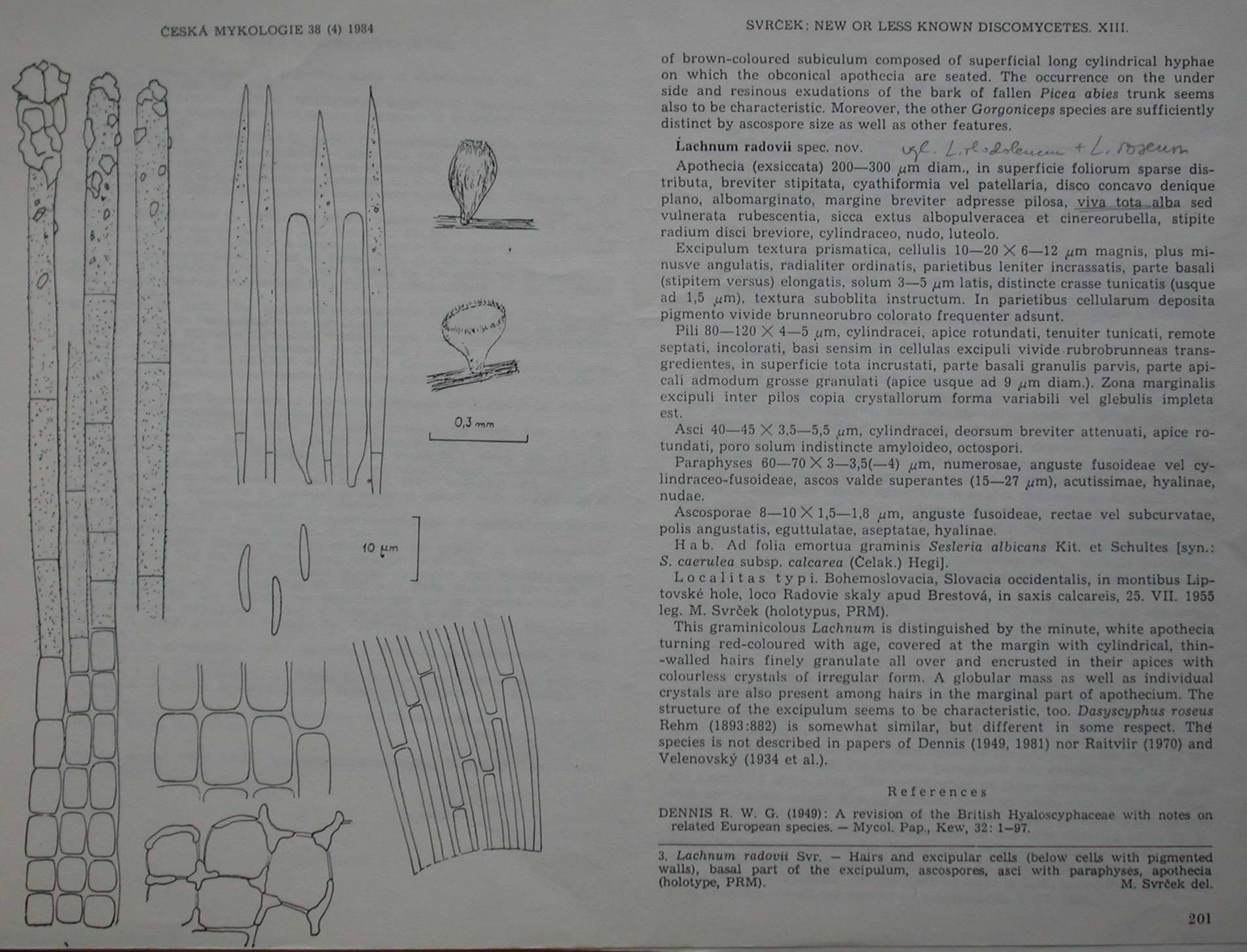 Svrcek-1984-New-or-less-13-3-0001.JPG
 Svrcek-1984-New-or-less-13-4-0001.JPG
I've compared these two short-spored Gorgonicepses (G. arctica, G. hypothallosa) with my specimen. Raitviir (2008) says there are only two short-spored wood-inhabiting species of Gorgoniceps. Still my specimen features don't coinside with previous descriptions.
There are major differences:
1) color of live apothecia: G. arctica described as "beige to pale ochraceous"; G. hypothallosa as "pale or dark gray, when dried brown to almost black, + turn red when mounted in KOH"; my specimen red when alive, reddish-brown in exsiccate, scarlet red in transmitted light and relises red pigment in water.
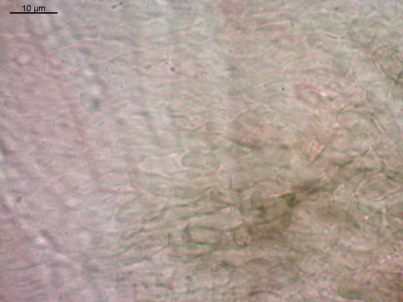
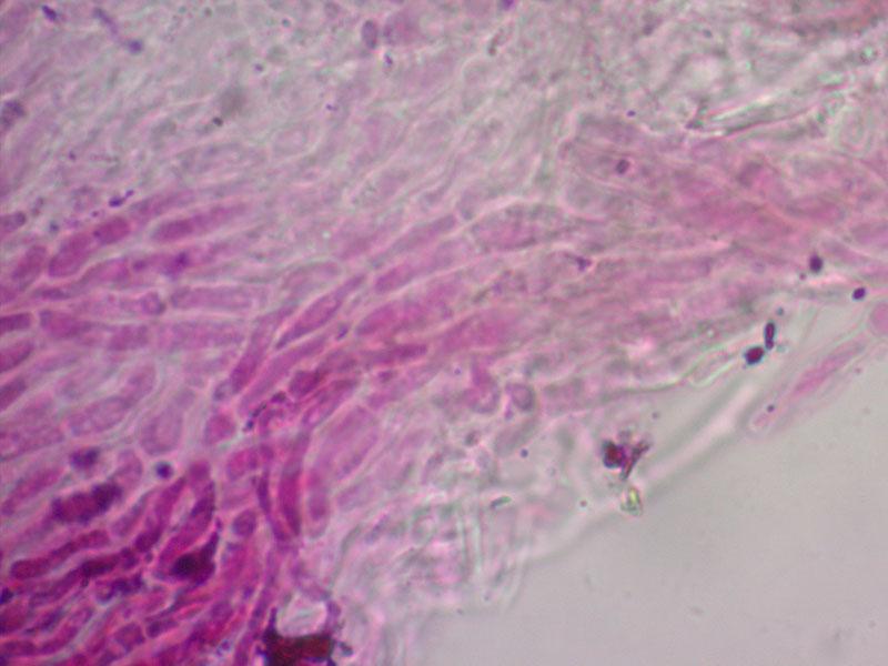
2) structure of excipulum: G. arctica has textura prismatica at base, cells to 12 mk broad, more narrow and clavate at the margin; G. hypothallosa has excipulum from parralel septate hypha 3-5 mk broad, with clavate cells at margin; my specimen textura oblita, from parralel hyphae 3-5 mk, with clavate ends in outer layer and at the margin. E.g. textura prismatica in G. arctica, and textura oblita in others two.
3) presence of subiculum: G. hypothallosa has developed subiculum from brown hyphae, G. arctica has not, neither my specimen. 4) habitat: G. arctica described from decaying stem of Alnus viridis (decidious wood); G. hypothallosa from bark of Picea abies, my specimen from bleached wood of Pinus sylvestris.
The structure of asci, paraphyses, spores seems coincide well in all three.
There are drowings of described species and my specimen, and comparative table. I would appreciate if you give me some instructions (i am only embarking to systematics, so have doubt what next). I had only one collection of this in last summer, and there are limited number of apothecia (about 20 at one wood piece). Probably, it should be observed more when it will summer again.
 short-spored-Gorgoniceps-compared-0001.xlsx
I agree with your comparison, but I am not sure if we can lay too much stress on colour. Perhaps there is a difference in the soulbility of the red-brown pigment, but such chemical characters are usually only found out when examining the type. And the colour may totally fade some ten years in the herbarium. Also the subiculum of hypothallosa should be examined - I hope it belongs to the fungus.
What is also important is to look for the croziers at the ascus base (in hypothallosa completely unclear), also to illustrate the ascus apex including apical ring at high resolution (perhaps always very similar but without an image we do not know).
The oblita is not very clear to me, on your drawing it is not seen (must have gel between the cells).
Finally, fresh material photographed and/or drawn in the living state would much help, especially concerning the spore droplets, septation of spores (already inside the living asci?), and contents of paraphyses.
Zotto
so i will look for another findings of this species to collect more vital information, and probably there will be some oppotunity to work with types in future.
"Textura oblita" i mean as in Ainsworth et al. (The fungi, 1973) (he does not say about the gel):
"..more or les parralel, agglutinated, thick-walled hyphae (textura oblita)"
I though (may be wrong) this term appropriate, since hyphae actually agglutinated and (?) thick-walled. Probably textura porrecta? ("more or less parralel, separable, thin-walled hyphae"). Or, should i read another source for tissue description?
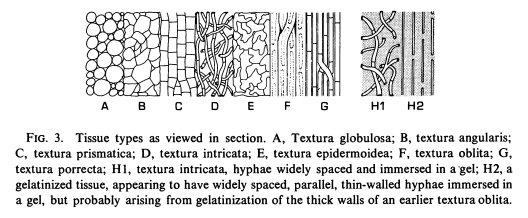
Durella has thick-walled cells too, but I am not sure if I would say oblita to this.
Zotto
There is pages from the paper (Raitviir, A., Huhtinen, S., 2002. A few out of many – interesting inoperculate, lignicolous discomycetes from Norway. Folia Cryptog. Estonica 39, 13–26.):