11-05-2026 12:32
 Bernard CLESSE
Pourriez-vous m'aider à identifier cette héloti
13-05-2026 15:26
 François Freléchoux
Bonjour,Voici une récolte faite il y a quelques j
12-05-2026 15:41
 Nicolas VAN VOOREN
Dear Ascolovers, especially interested in Pezizale
13-05-2026 12:05
 Thierry Blondelle
Bonjour à tous,J'aimerais avoir confirmation de c
10-05-2026 23:17
 Andreas Gminder
Hello,today we found in a moist steep decidous for
28-04-2026 20:07
 Lothar Krieglsteiner
... on twig in the air at standing Ceratonia siliq
27-04-2026 20:52
Lothar Krieglsteiner
Found on hanging tiwg of Olea europaea in dried-ou
11-05-2026 20:22
Lothar Krieglsteiner
on attached twig of standing Ficus caricaquite uns
29-04-2026 10:44
Lothar Krieglsteiner
growing at moist, drying-out soil at the side of a
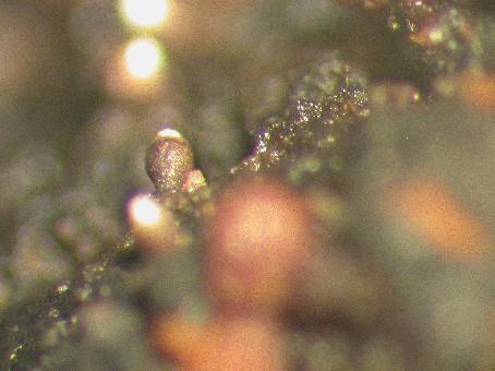
It occurs on exudate of wounds of Prunus pensylvanica in PEI and NB Canada.
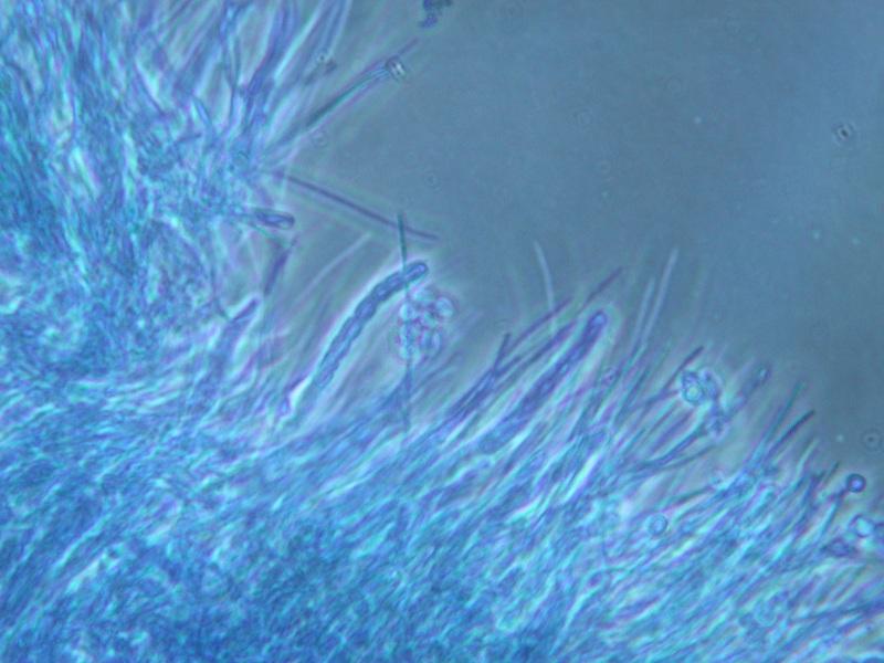
The ascomata are about 200-400 um tall and 150-200 um wide, and urceolate in shape. The walls are light brown and become melanized as it matures. Internally, the walls consist of linear, periclinally arranged hyphae. The asci are narrowly cylindric (c. 35 x 4 um), and lack an evident apical apparatus. They break down at maturity, producing a dry mass of spores that collects around the ostiole, and within the cavity in the upper part of the ascoma. Externally, the spore mass appears white to very pale yellow. The ascospores are uniseriately arranged, 8 per ascus, colourless, unornamented, and c. 3-4 x 2-3 um. They are ellipsoidal, but slightly compressed on the long axis.