
28-04-2026 20:07
 Lothar Krieglsteiner
... on twig in the air at standing Ceratonia siliq
14-04-2026 05:32
Ethan CrensonHi all, A few weeks back a friend pointed out som
28-04-2026 20:33
Vitus SchäfftleinHello, I found Trochila ilicina on Ilex aquifoliu

30-04-2026 10:28
Rot BojanHello, by appearance I would say that I am dealing
27-04-2026 18:48
Tony MoverleyCollected 23rd April 2026, Norfolk, EnglandSwarms
27-04-2026 20:52
Lothar Krieglsteiner
Found on hanging tiwg of Olea europaea in dried-ou
28-04-2026 22:51
 Bernard CLESSE
Bonsoir à toutes et tous,Pourriez-vous m'aider à
29-04-2026 08:01
Lothar Krieglsteiner
... on twig attached to small tree of Citrus auran
29-04-2026 10:44
Lothar Krieglsteiner
growing at moist, drying-out soil at the side of a
Pyrenomycet with brown, two celled spores
Gernot Friebes,
21-04-2010 17:08
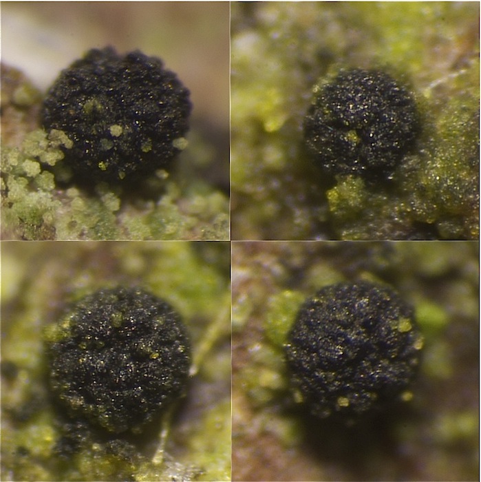
here I'd need help because I have never seen a pyrenomycet with a combination of these characters. It is a pyrenomycet which I found twice: the first time on 19/2/2010 and the second time four days ago. Both times it grew on the bark of living Salix, some 150 cm above the ground. The first collection was immature and had only hyaline spores while the second collection had brown, two celled spores. However, I observed asci only in the first collection while in the second collection there were only spores.
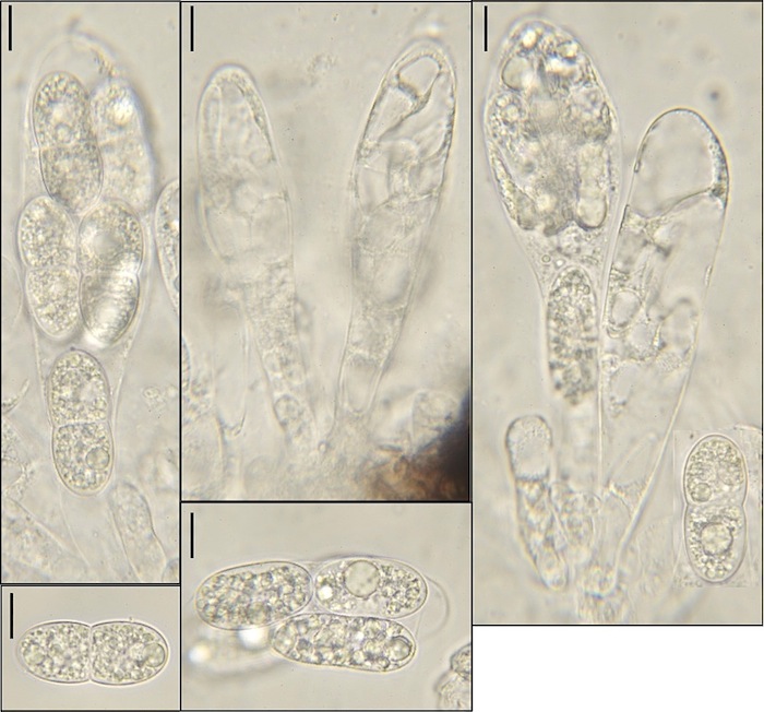
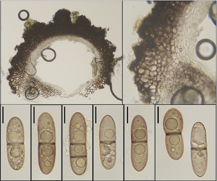
I am quite sure that these two collections are the same species because they had exactly the same ecology and macroscopical appearance. Furthermore, the immature spores of the second collection looked like the spores of the first collection. The asci (of the first collection) were up to 120 x 30 µm, unitunicate and clavate, without a visible apical apparatus and 8-spored. The spores were always two celled except of one spore with two septa, primarily hyaline, then brown with a darker septum, 32-37,5 x 9,5-12(14) µm. The ascomata were free, rough, black and very soft when wet.
Best wishes and many thanks
Gernot
Here is a picture of the perithecia (of the first collection but they actually looked the same):
Gernot Friebes,
21-04-2010 17:08
Re:Pyrenomycet with brown, two celled spores
micros of collection one (scale=10 µm):
Gernot Friebes,
21-04-2010 17:09
Re:Pyrenomycet with brown, two celled spores
micros of collection two (scale=10 µm):
Jacques Fournier,
21-04-2010 21:09

Re:Pyrenomycet with brown, two celled spores
Hi Gernot,
again a puzzling collection, do you ever find ordinary fungi?
I definitely have no idea to accommodate it. Maybe some further characters would help. The ascomata seem to be ostiolate, with some tubercles on top, can you confirm ? How big are the ascomata, and do they turn cupulate on drying or remain globose?
On your photos, asci seem short-stipitate, is it trus? Did you notice any paraphyses among asci?
I guess the occurrence on living branches makes it very different from usual saprobic pyrenomycetes. I hope somebody else will be more helpful.
Best wishes,
Jacques
Gernot Friebes,
21-04-2010 21:41
Re:Pyrenomycet with brown, two celled spores
Hi Jacques,
thank you for your answer!
"again a puzzling collection, do you ever find ordinary fungi?"
Sorry :)
I didn't see ostioles in both collections so I can't tell you. But it is true that the fungus has a very rough, Bertia-like surface with a very soft, non-carbonized wall. The ascomata have a diameter of some 300-500 µm and they remain globose when dry (but they turn quite stiff). I would consider the asci short-stipitate, yes, and I didn't see any paraphyses. I think you misunderstood the ecology because my fungus grew on the bark of living Salix, where you would also look for Hysterium and related fungi.
Best wishes,
Gernot