02-12-2017 11:41
Thorben HülsewigHi there,a few days ago i found this fungus next t
30-11-2017 23:31
Garcia SusanaPeritecios en torno a 300-400 um de diámetro con
01-12-2017 11:35
José Antonio López-SáezWould you be so kind to help me identify the follo
01-12-2017 07:36
 Nicolas VAN VOOREN
Bonjour.Un nouveau numéro des Cahiers de la FMBDS
29-11-2017 22:39
F. JAVIER BALDA JAUREGUIuna idea ,para este anamorfo que crecia sobre Pyth


27-11-2017 14:04
Eduard OsieckThis Hilberina-like pyrenomycete was found on a de
26-11-2017 15:01
Eduard OsieckWould anyone be able to confirm that this anamorph
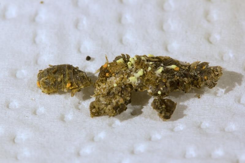
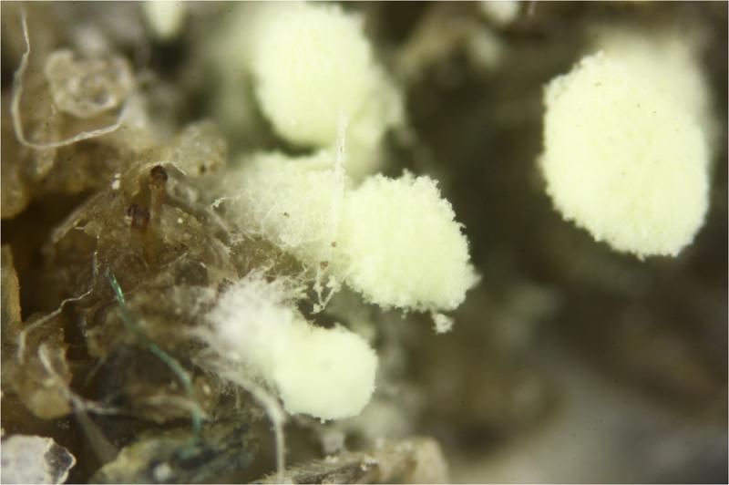
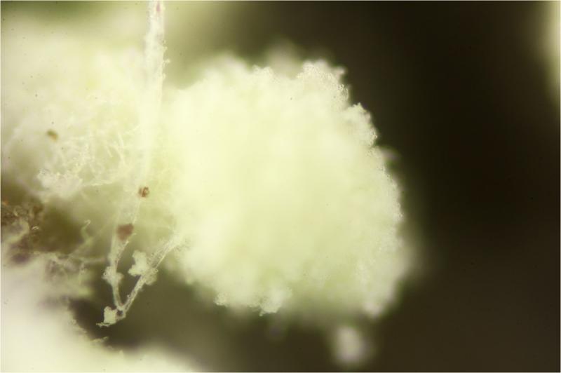
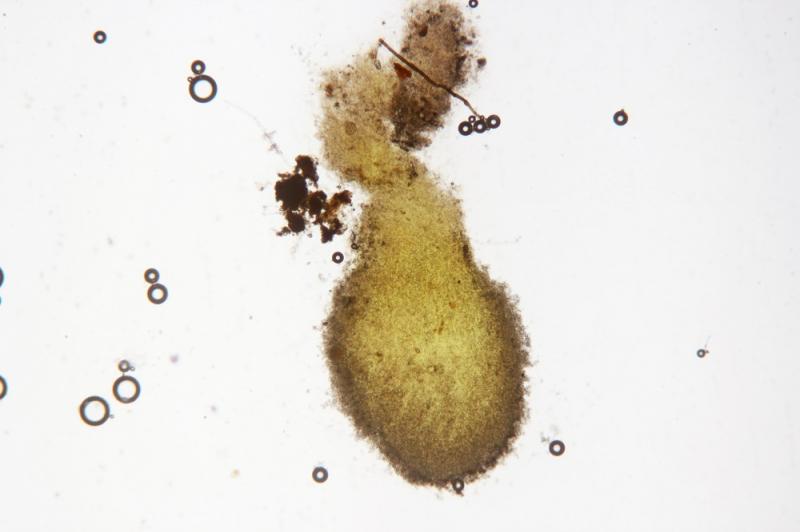
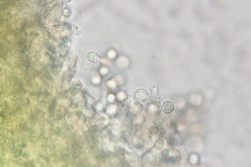
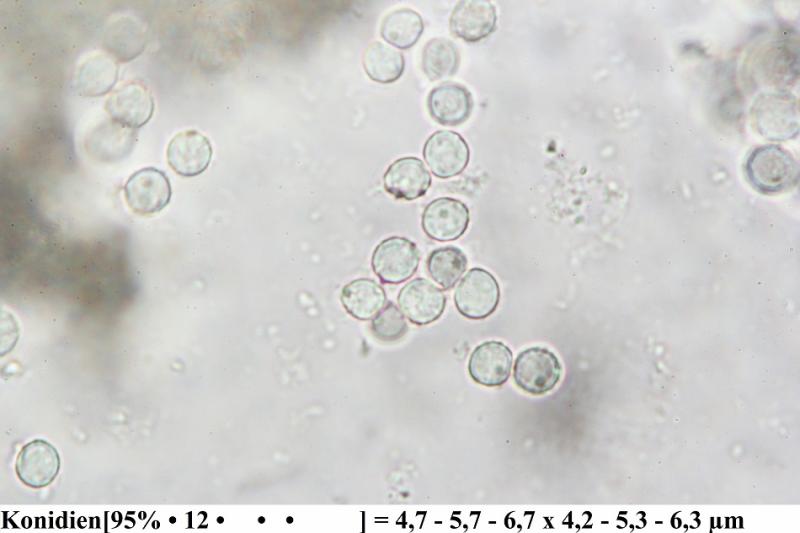
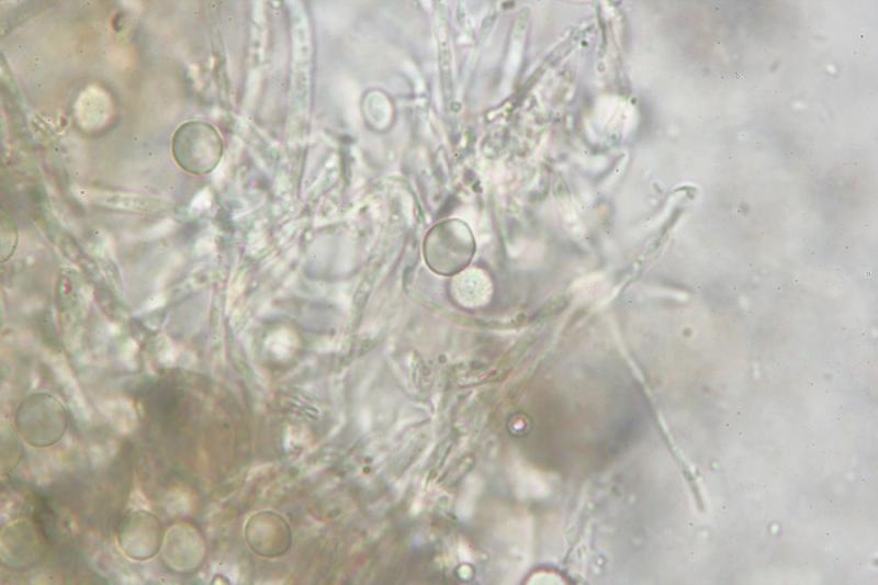
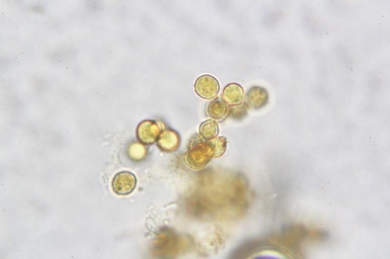
Chrysosporium sp. ? on Porcellio scaber
Thorben Hülsewig,
02-12-2017 11:41
a few days ago i found this fungus next to Gymnoascus devroeyi.
The conidie are verrucose and become orange in Lugol.
Could that be a Chrysosporium ?
thanks in advance,
Thorben