
16-01-2018 08:53
 Zoltan Lukacs
Hello, I've picture only. Very little konidiospore
12-01-2018 21:27
 Claude Kaufholtz-Couture
Bonjour à tous, Je récidive pour la description

13-01-2018 13:46
Claude Kaufholtz-Couture
Bonjour à tous,J'ai deux autres collections de Sc
14-01-2018 10:32
 Castillo Joseba
Me mandan el material seco... en hoja de quercusA
06-01-2018 20:08
 M Jonathan
Bonjours, j'espère que quelqu'un peut m'aider ave
12-01-2018 23:43
 Bernard CLESSE
Bonsoir à toutes et tous,Un ami me confie cette r
12-01-2018 20:56
 Vasileios Kaounas
Found in 08-01-18, in in a rotten section of Aspho
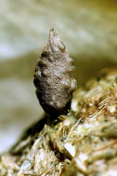
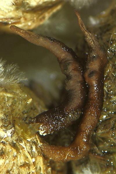
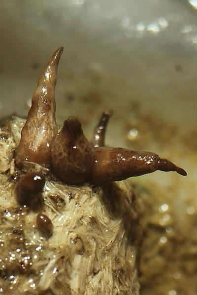
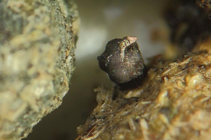
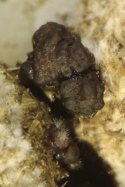
Xylaria ? on rubbit dung
Zoltan Lukacs,
16-01-2018 08:53
Hello, I've picture only. Very little konidiospores. Any idea?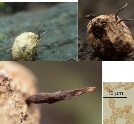
Peter Püwert,
16-01-2018 10:29

Re : Xylaria ? on rubbit dung
Hi,
see the avatar, in my opinion is this Podosordaria/ Xylaria tulasnei, mostly on rabbit dung.
Greetings Peter.
see the avatar, in my opinion is this Podosordaria/ Xylaria tulasnei, mostly on rabbit dung.
Greetings Peter.
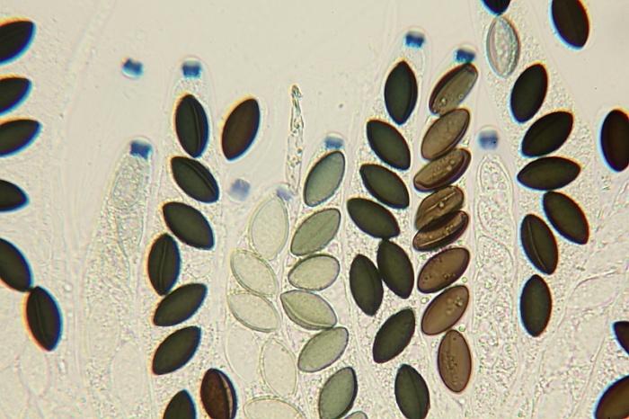
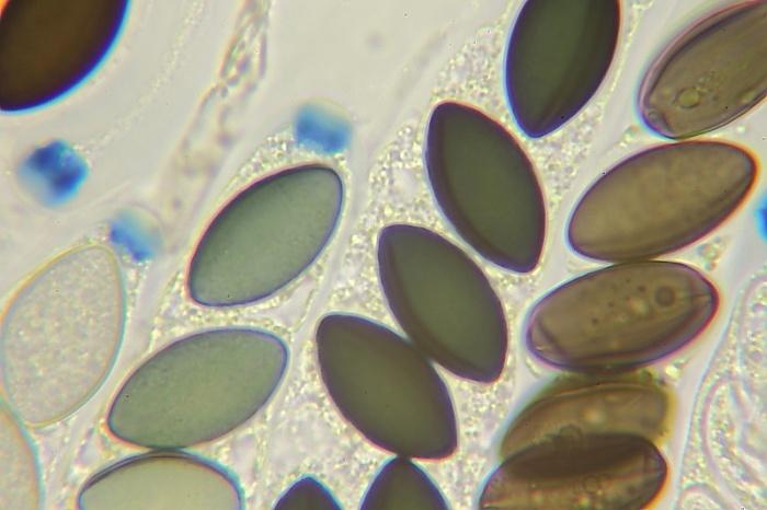
Zoltan Lukacs,
16-01-2018 11:38
Re : Xylaria ? on rubbit dung
Hi Peter,
It seems a good solution.
Many thanks
Zoltan
It seems a good solution.
Many thanks
Zoltan