10-03-2013 11:10
 Björn Wergen
Hi again,do you also think that this collection ca
07-05-2013 11:54
 Miguel Ángel Ribes
Another Mollisia? in wood without bark, no more th
06-05-2013 10:55
 Yannick Mourgues
Bonjour à tous.Qui aurait ces docs en pdf ? Ou qu
04-05-2013 22:20
 Chris Yeates
Bonsoir tousthere seems to have been a glut of cop
05-05-2013 14:25
Stefan BlaserHello everybody Here's already my next problem:
06-05-2013 00:07
 Michel Hairaud
Bonjour, Je sollicite votre aide pour cette réco

05-05-2013 11:13
 Nicolas VAN VOOREN
Bonjour.Je cherche l'article suivant : Yao Y.J. &a
Trematosphaeria pertusa
Björn Wergen,
10-03-2013 11:10
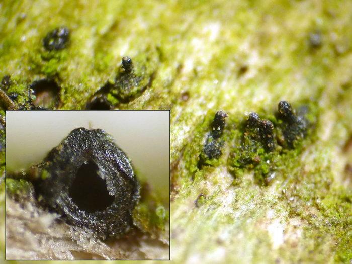
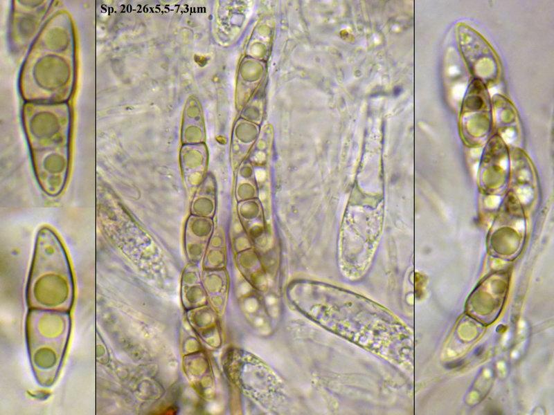
Hi again,do you also think that this collection can be T. pertusa? I have found several articles and photos of this species but because of several contrarieties I do not really know how T. pertusa should look like. The spores of this collection are hyaline and 1septate in immature state, but they become 3 septate and pale greenish to greenbrown.
Macroscopically the perithecia are about 0,4-0,7 mm broad and sphaerical, with a pointed Ostiolus and covered with brown hairs (?).
Collected on attached twigs of Tilia cordata.
regards,
björn
Yannick Mourgues,
10-03-2013 11:39
Re : Trematosphaeria pertusa
Hi Björn .
I think it's not T. pertusa because in this specie (see Zhang & al 2008, Are Melanomma Pulvis-pyrius and Trematosphaeria pertusa congeneric ? - Fungal Diversity 33 : 47-60) spores are larger and longer, and dark brown :
"27.5-32.5x7,5-8,5 , fusiform with broadly to narrowly rounded end, dark brown, 1(-3) septate, (...), smooth to finely verruculose, without gel sheat"
Have your spores a gel sheat ?
May be you should make a vertical section of peridium to see cells structure.
Have you looked for in Massarina or Herpotrichia (subiculum ?) species ?
Yannick
Björn Wergen,
10-03-2013 11:56
Re : Trematosphaeria pertusa
Hi Yannick,
Herpotrichia is a good idea. But it is not H. herpotrichoides since I had this species already on Rubus sp. with different characters. I had also look for Massarina but I did not find any matching species.
I did not see a gel coat surrounding the spores.
regards,
björn
Herpotrichia is a good idea. But it is not H. herpotrichoides since I had this species already on Rubus sp. with different characters. I had also look for Massarina but I did not find any matching species.
I did not see a gel coat surrounding the spores.
regards,
björn
Björn Wergen,
10-03-2013 14:25
Re : Trematosphaeria pertusa
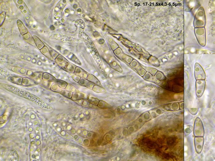
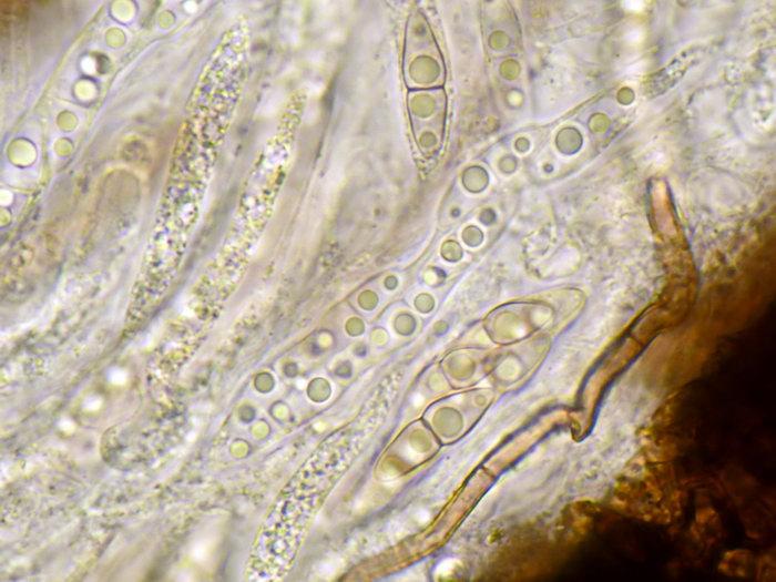
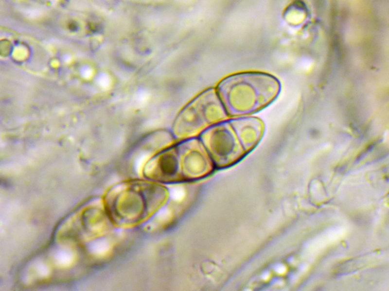
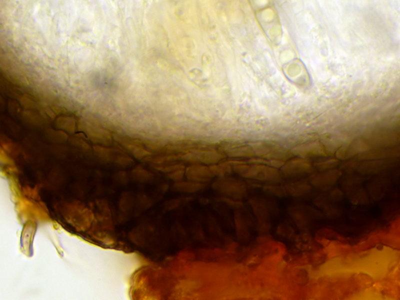
Here are some more data, especially a cut through a perithecia.
Some of them have longer Ostioles and are crowded together. I have seen no gel coat, also not with Indian Ink.
Wall is 25-32µm thick.
regards,
björn
Some of them have longer Ostioles and are crowded together. I have seen no gel coat, also not with Indian Ink.
Wall is 25-32µm thick.
regards,
björn
Ying Ying Zhang,
11-03-2013 03:13
Re : Trematosphaeria pertusa
??Hi Bjorn,
This species do not fit T. pertusa well. The ascospore of T. pertusa is pigmented. The ascomata do not covered with anything ("naked"). The ostiole (opening) is wide. But the shape of ascospores and asci of this collection are very similar with T. pertusa. I would like to take it as another species of Trematosphaeria s. s. As Trematosphaeria seems not rare in Europe, thus close relatives might also not rare.
Cheer up!
Ying
Björn Wergen,
11-03-2013 21:40
Re : Trematosphaeria pertusa
Hi Ying,
thanks for the respond. I have recently found this species again, on Ulmus.
regards,
björn
thanks for the respond. I have recently found this species again, on Ulmus.
regards,
björn
Björn Wergen,
08-05-2013 16:44
Re : Trematosphaeria pertusa
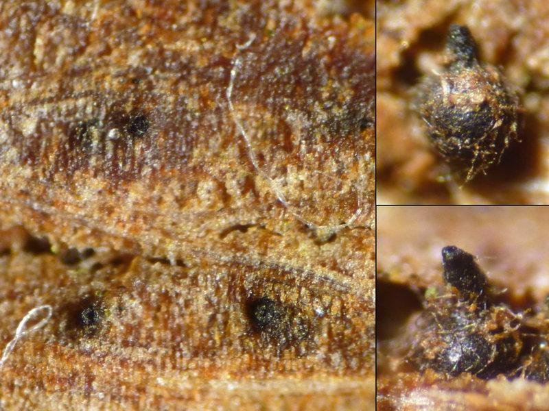
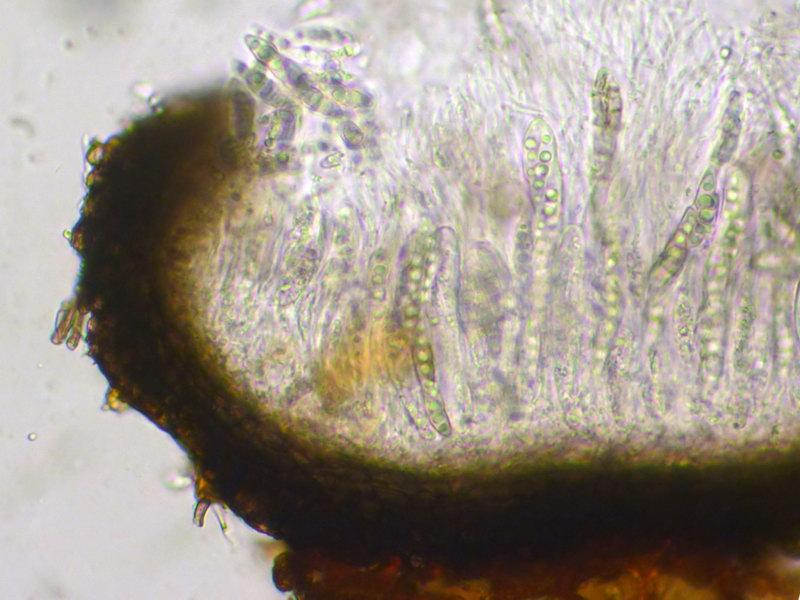
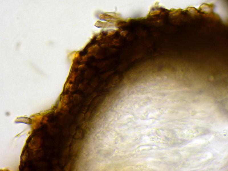
I have found it again, this time on decorticated Acer stems (~5cm).
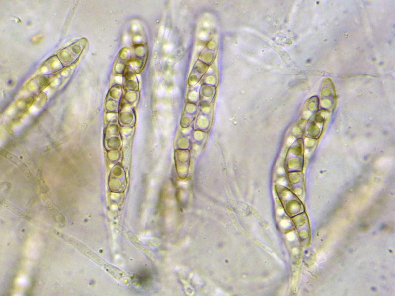
The spores are a bit smaller than in the other 2 collections, and I haven't seen any spores with more than 1 septa.
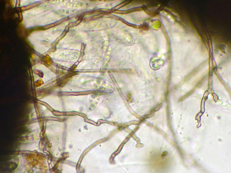
As already seen in the other photos, this collection also had a hyphal layer on the small, black perithecia (diam. ~ 0,6mm, beaked).
I have called it "Trematosphaeria lignicola" for now.
I will add it to my special herbarium for further studies.
regards,
björn
The spores are a bit smaller than in the other 2 collections, and I haven't seen any spores with more than 1 septa.
As already seen in the other photos, this collection also had a hyphal layer on the small, black perithecia (diam. ~ 0,6mm, beaked).
I have called it "Trematosphaeria lignicola" for now.
I will add it to my special herbarium for further studies.
regards,
björn