31-12-2013 01:28
 Rubén Martínez-Gil
Hola a todos.El pasado sábado encontramos esta Ot
28-12-2013 23:24
 Michel Hairaud
Bonsoir à tous, Hi to everyone,Pour la 2eme anné
28-12-2013 01:00
Rubén Martínez-Gil
Hola a todos.Pongo unas fotos de este asco que en
26-12-2013 19:59
 Enrique Rubio
These very small (up 70 125 microns), superficial,
26-12-2013 20:37
Eduard OsieckPiece of decorticated Fraxinus wood, collected 4 m
25-12-2013 22:52
Rubén Martínez-Gil
Hola a todos.Pongo fotos de una recolecta sobre ma
Otidea concinna?
Rubén Martínez-Gil,
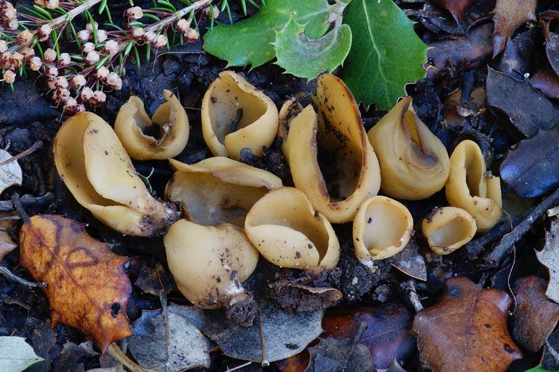
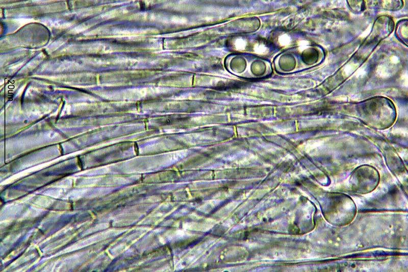
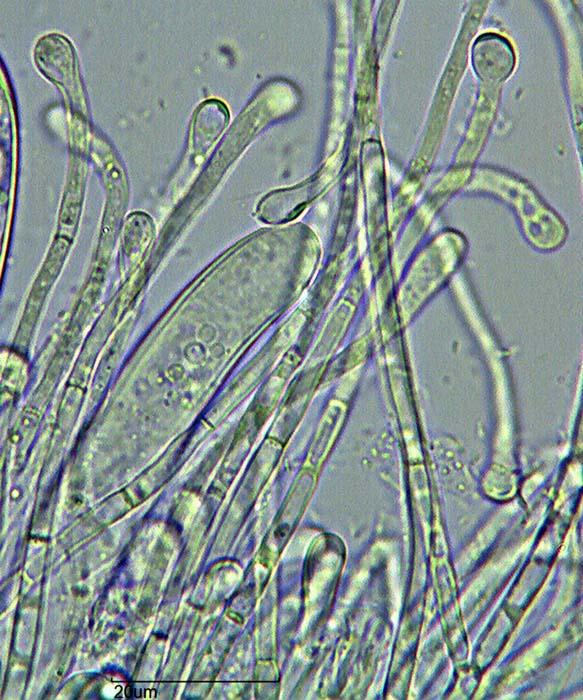
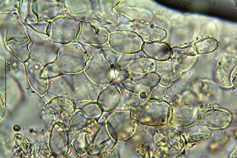
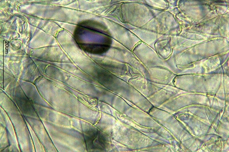
31-12-2013 01:28
Hola a todos.El pasado sábado encontramos esta Otidea en suelo de encinar ácido y en una zona húmeda.
Tamaño de 1 a 4 cm de altura.
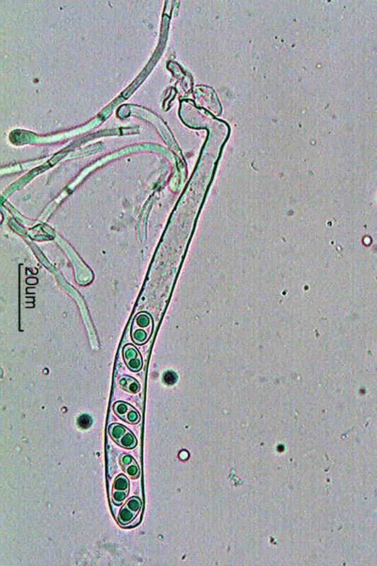
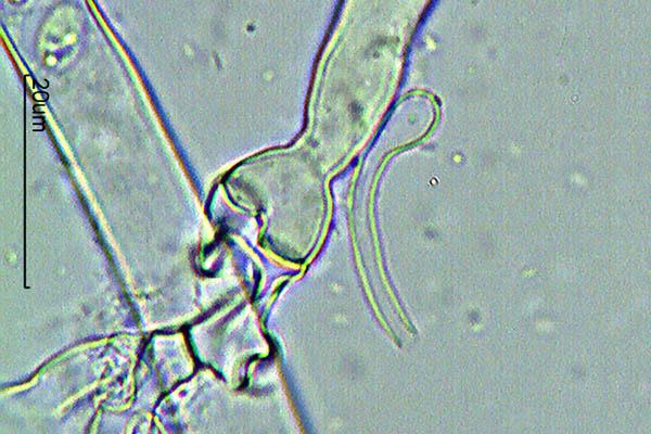
Paráfisis septadas, algunas bifurcadas y engrosadas en el ápice con alguno de ellos curvo o en forma de cayado.
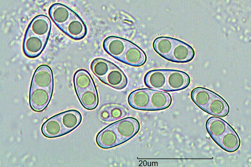
Esporas de 10,7-13,1 x 5,2-6,1 micras.
Creo que podría tratarse de O. concinna, ¿Qué os parece?
Gracias a todos
Un saludo
Rubén