
16-03-2018 09:37
Gilbert MOYNEHabitat : sur rameau mort aérien de gui (Viscum a
20-01-2020 18:55
 Andgelo Mombert
Bonsoir, Un petit discomycète vert noirâtre mes


21-02-2020 14:47
Dragiša SavicHi to all, I need help identifying this lichenicol
21-02-2020 11:48
 Michel Hairaud
Bonjour / Hi to everyone Le 1er numéro 2020 d'As
14-02-2020 18:19
 Charles Aron
Hello everyone. My first contribution to the forum
Diplodia?
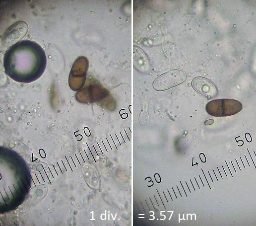
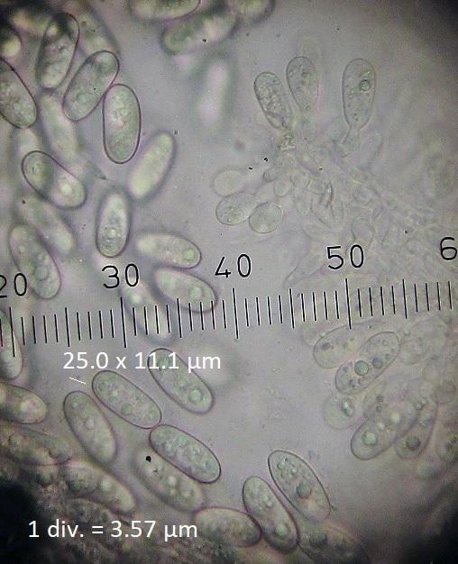
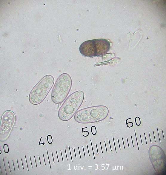
Riet van Oosten,
23-02-2020 18:10

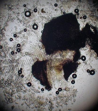
Hello,
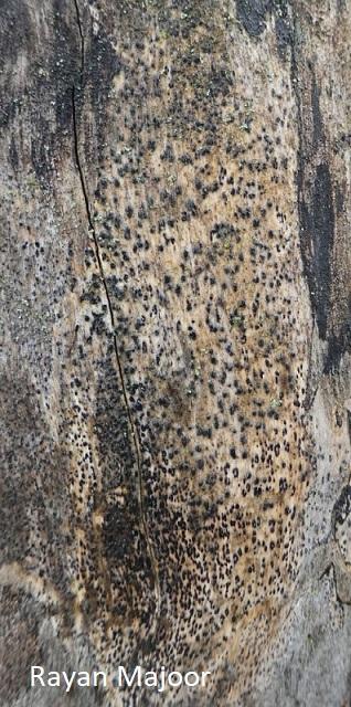
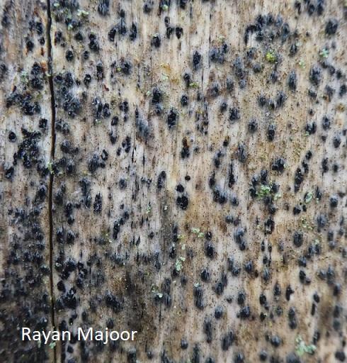
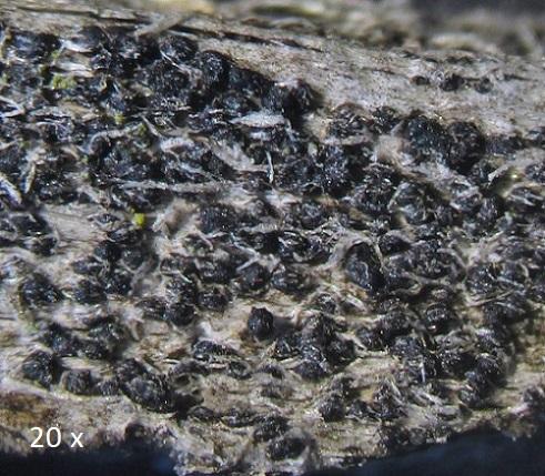
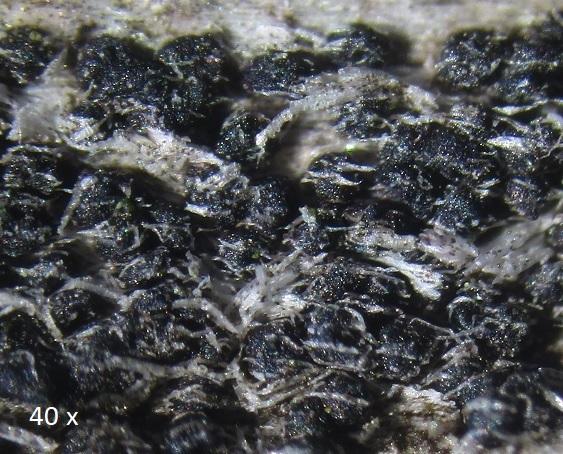
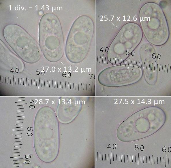
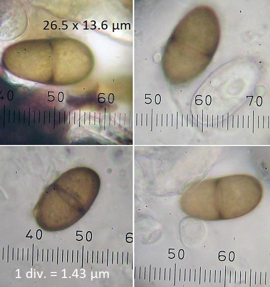
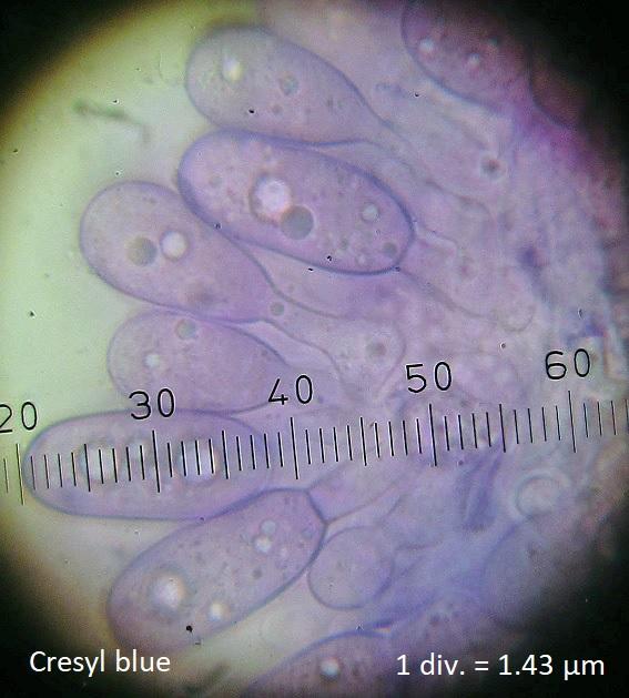
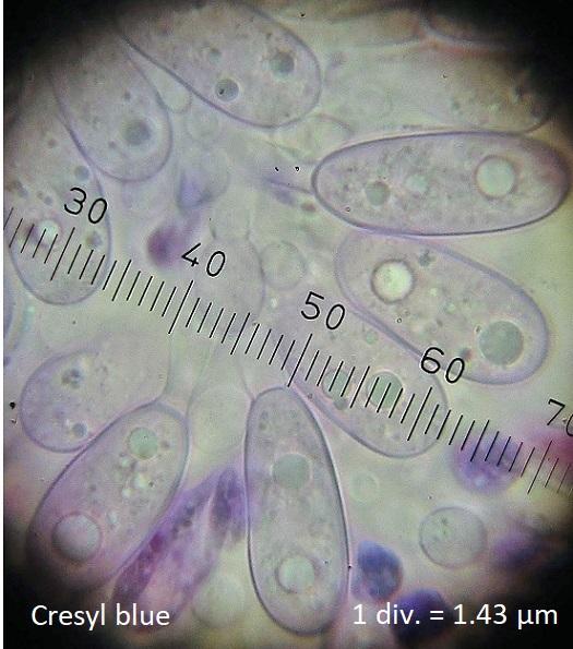
Found by Rayan Majoor, Jan. 2020, The Netherlands.
With the help of the publication mentioned in this topic (thank you Angel and Josep!) I think it's a Diplodia
Key to the genera, page 66.
Key to Diplodia, page 82.
1-2-5-6 ..... don't know the host (I will ask Rayan)
Spore sizes average < 28 µm
Only a few brown 1-septate spores.
Greetings, Riet

Angel Pintos,
23-02-2020 19:05

Re : Diplodia?
Hi Riet,
following the keys of the article you mention it is a Diplodia, following the measures you give can be D. africana.
I had a similar one on Celtis australis, everything indicated that it was a Diplodia sp. according to the article and it turned out to be a Sardiniella sp.
Good luck
Regaards
Angel
following the keys of the article you mention it is a Diplodia, following the measures you give can be D. africana.
I had a similar one on Celtis australis, everything indicated that it was a Diplodia sp. according to the article and it turned out to be a Sardiniella sp.
Good luck
Regaards
Angel
Riet van Oosten,
23-02-2020 19:13
Re : Diplodia?
Thank you very much for your help Angel!