
26-10-2020 09:58
 Nicolas VAN VOOREN
Hi.Here is the tiny white discomycete (~0.1 mm Ø)
27-10-2020 21:40
 Joop van der Lee
There is some doubt in how we address this species
27-10-2020 13:15
 Viktorie Halasu
Hello,would anyone has this paper please? I'd like
23-10-2020 12:57
 Marek Wolkowycki
Hello Bia?owie?a Primeval Forest on live shoots o
26-10-2020 07:48
 Blasco Rafael
Hola, esta muestra estaba sobre tallos de Angelica
25-10-2020 12:35
Juuso ÄikäsThese were growing on a low branch of a deciduous
24-10-2020 14:28
 Castillo Joseba
De esta mañana recolectada en acicullas de abet
Tiny white Helotiales
Nicolas VAN VOOREN,
26-10-2020 09:58
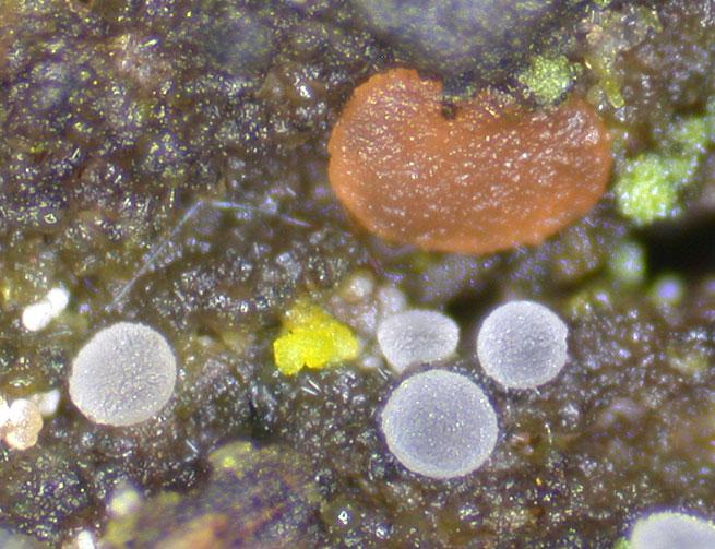
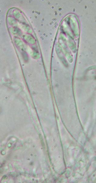
Hi.Here is the tiny white discomycete (~0.1 mm Ø) growing on dead branch of Salix (still attached), with Orbilia vinosa.
Asci 72-76 x 10 µm, without crozier, IKI-
Paraphyses filiform, 1.5-2 µm Ø, very flexuous.
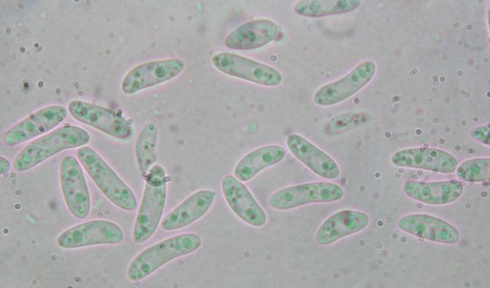
Ascospores of heterogeneous shape, 10-15 x 3.5-4 µm
I have no idea of the genus...
Nicolas
Stip Helleman,
27-10-2020 12:26

Re : Tiny white Helotiales
Hi Nicolas
Are you sure no hairs? They can be very inconspicuous in Hyaloscypha in my experience.
Cheers
Stip
Are you sure no hairs? They can be very inconspicuous in Hyaloscypha in my experience.
Cheers
Stip
Nicolas VAN VOOREN,
27-10-2020 12:47
Re : Tiny white Helotiales
I can try a new mount to check that.
Adam Polhorský,
27-10-2020 15:41
Re : Tiny white Helotiales
Hello,
Hyaloscypha minuta seems like good option. The hairs are not well developed there.
Nicolas VAN VOOREN,
27-10-2020 17:39
Re : Tiny white Helotiales
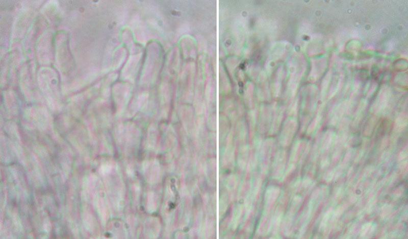
Here are two pictures of marginal cells (taken from the outside of an apothecium). No hairs or no hairs of Hyaloscypha-type.
Stip Helleman,
27-10-2020 19:13
Re : Tiny white Helotiales
How disappointing, I would have bet on it.
With this lack of characters I have no idea for a genus.
Cheers, Stip
With this lack of characters I have no idea for a genus.
Cheers, Stip
Hans-Otto Baral,
28-10-2020 06:40

Re : Tiny white Helotiales
I agree with H. minuta (= Parorbiliopsis minuta). That species has very minute hairs or not at all any.
But I estimate the diameter of the apos at max. 0.5 mm.
Zotto
Nicolas VAN VOOREN,
28-10-2020 10:47
Re : Tiny white Helotiales
OK thanks.