
09-12-2012 20:42
 Björn Wergen
Hi all,I have found this small pyrenomycete on dea
28-10-2012 09:54
Björn Wergen
hello everybody,this is the second time I have fou
08-12-2012 11:44
Björn Wergen
Hello everybody,I am back with some new, unclear p
04-12-2012 23:14
 Christiane Baethcke
Dear friends, I need your help again, this time w
06-12-2012 21:42
Salvador TelloDe hasta 5 mm en tierra con mucho estiercol de ove
06-12-2012 20:25
 Alessio Pierotti
Help ! I search this work: Kobayasi, 1960. - Ascos
05-12-2012 21:21
 Gilles Corriol
Bonjour à tous,Nouvellement inscrit au forum Asco
05-12-2012 17:15
Salvador TelloHola a todos.Pienso que este hongo puede ser Hypoc
Pocillum cesatii ?
Camporesi Erio,
09-12-2012 20:31
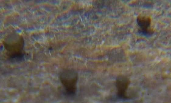
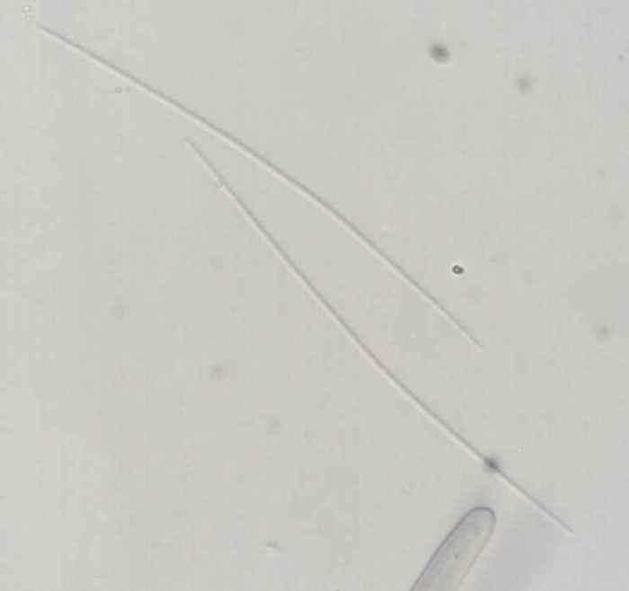
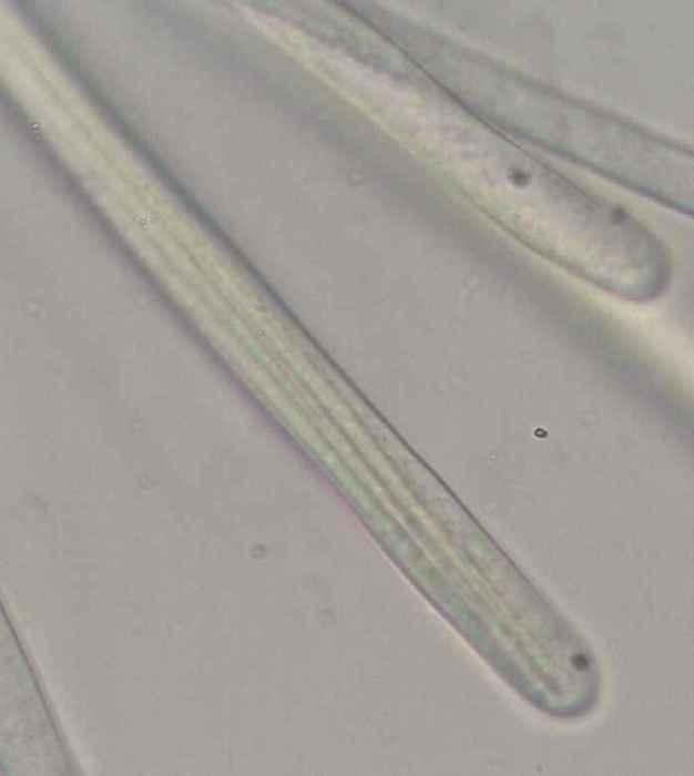
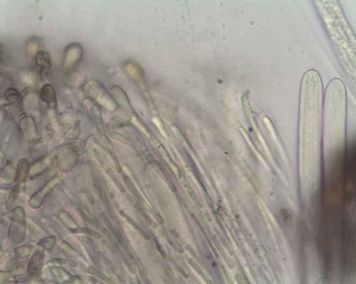
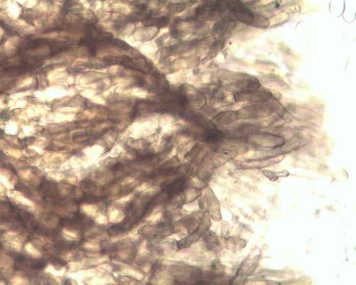
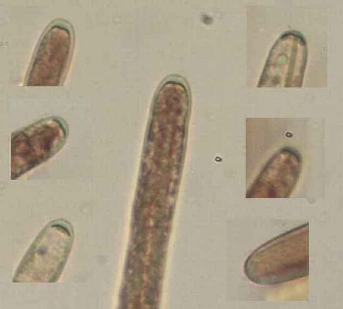
is this fungus Pocillum cesatii ?
Apothecia up to 0,5 mm in diameter.
Ascospores up to 140 x 1 micron .
Asci IKI-
On dead and land leaf of Quercus , April 2012.
Ciao
Erio
Hans-Otto Baral,
09-12-2012 21:01

Re : Pocillum cesatii ?
Hi Erio
yes, for sure! A quite rare species apparently.
Very well seen on your photo with IKI is the nasse apicale in the ascus apex, also known from typical Vibrissea species.
When did you collect this fungus? In summer?
Zotto
yes, for sure! A quite rare species apparently.
Very well seen on your photo with IKI is the nasse apicale in the ascus apex, also known from typical Vibrissea species.
When did you collect this fungus? In summer?
Zotto
Camporesi Erio,
09-12-2012 21:28
Re : Pocillum cesatii ?
Hi Zotto,
I collected it the last spring (25 Aprill 2012 ..... 700 metres of altitude about... after copious rains)) in a deciduous forest (Quercus , Ostrya and so on...).
Ciao
Erio
I collected it the last spring (25 Aprill 2012 ..... 700 metres of altitude about... after copious rains)) in a deciduous forest (Quercus , Ostrya and so on...).
Ciao
Erio
Hans-Otto Baral,
09-12-2012 21:32
Re : Pocillum cesatii ?
Ah, you wrote the month already :-)
Interesting species, perhaps adapted to heavy rainfall in spring? I assume it was not on the bank of a rivulet, like Vibrissea?
Interesting species, perhaps adapted to heavy rainfall in spring? I assume it was not on the bank of a rivulet, like Vibrissea?
Camporesi Erio,
09-12-2012 23:12
Re : Pocillum cesatii ?
Yes Zotto,
the leaf was in a humid and shadowy place, but not on the bank of a rivulet.
Ciao
Erio
the leaf was in a humid and shadowy place, but not on the bank of a rivulet.
Ciao
Erio