
12-06-2026 14:50
 François Freléchoux
Bonjour, Voici la brève description d'une Mollis
10-06-2026 21:16
François Freléchoux
Bonsoir,Le dernier du jour, en attendant votre avi
11-06-2026 19:01
 William Slosse
Hello all,In an attempt to make a culture of a sus
11-06-2026 19:03
 Nicolas VAN VOOREN
Chers membres d'Ascofrance,Le site sera placé en

09-06-2026 18:32
Camille MertensSur morceau de roseau immergé 0,5 - 0,7 mm de dia
10-06-2026 12:54
Steve ClementsBonjour encore, Pouvez-vous m'aider, s'il vous pl
10-06-2026 21:07
François Freléchoux
Toutes les tiges de gentianes jaunes de l'an pass�
10-06-2026 13:41
François Freléchoux
Bonjour à nouveau, Voici une trouvaille d'hier.

Hello, dear friends!
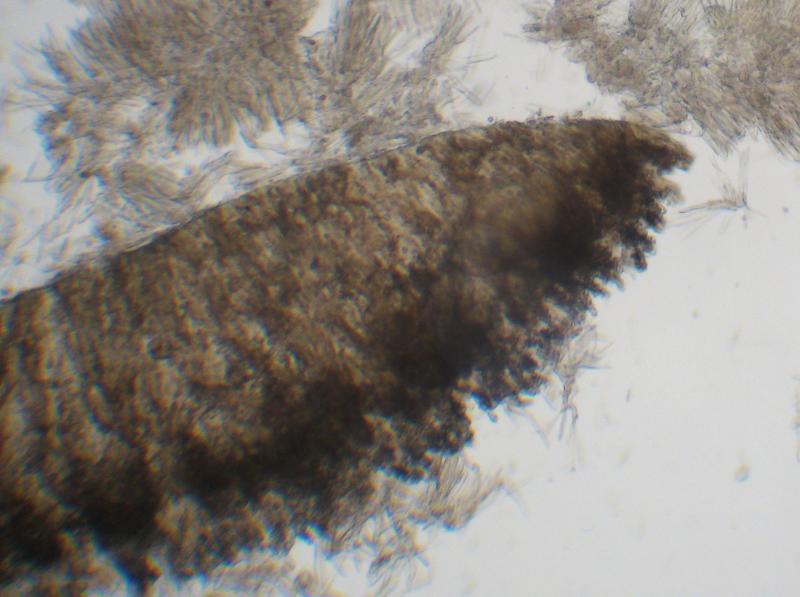
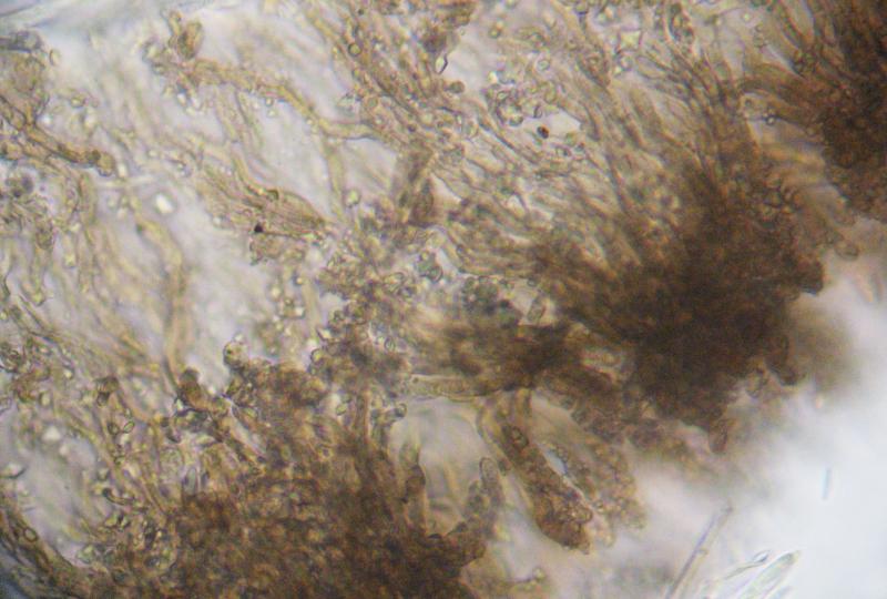
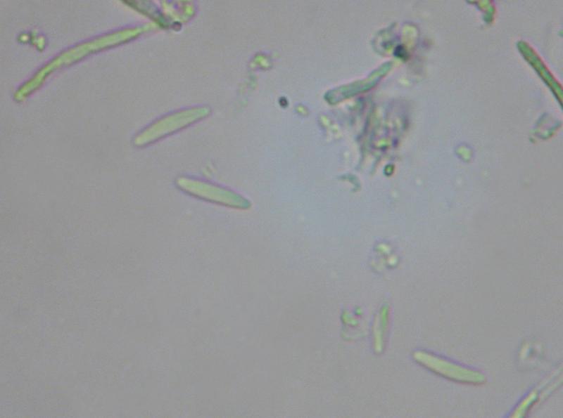
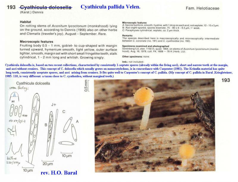
These 2 specimens some time ago i identidied as C. cyathoidea. Now I see some differences in spore morphology, and I wonder whether one of them could be C. pallida.
The 1st was examined in fresh condition, the 2d in exciccated state.
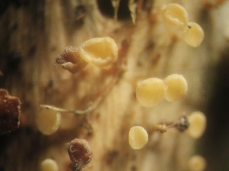
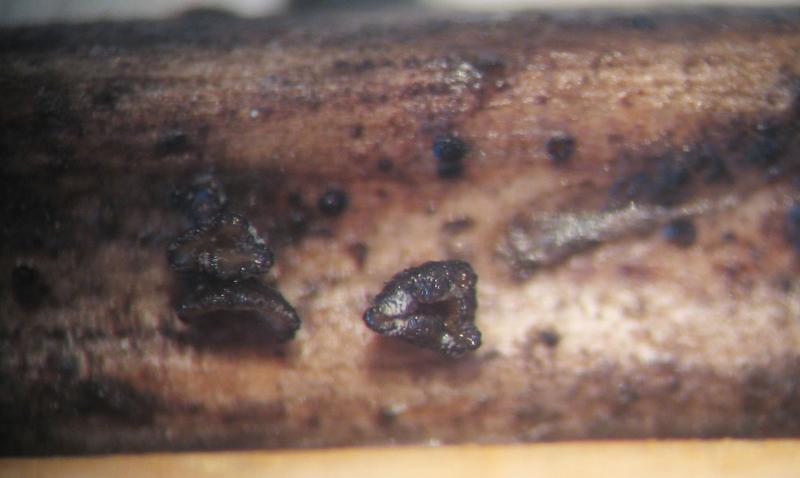
So, the 1st specimen was collected in oak forest, on Urtica dioica rotten stem.
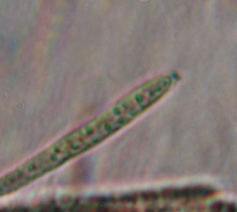
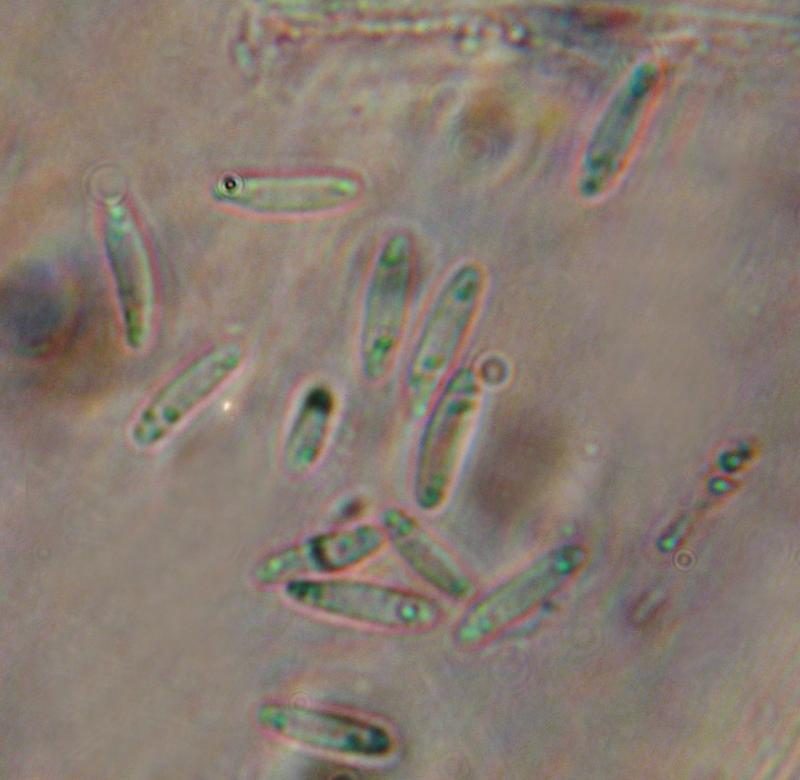
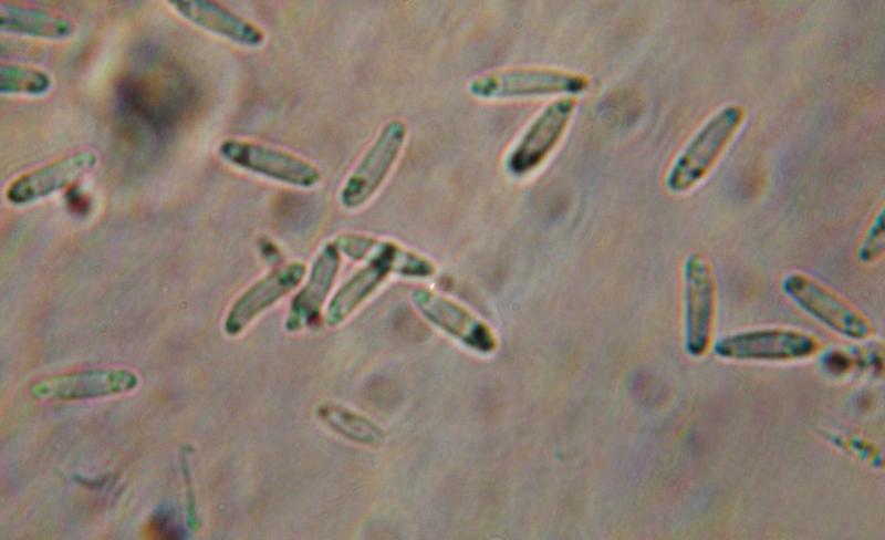
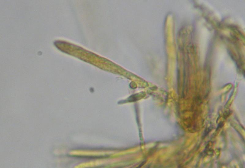
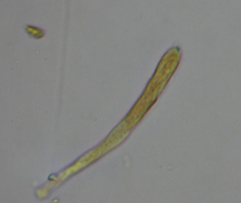
Spores 7,3-12,6*2,2-3,6 um, with 1-3 small oil drops on each end.
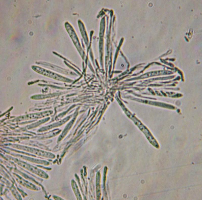
Asci IKI B, with croziers, 49-68*3,6-5,5 um
Cheers,
Irina
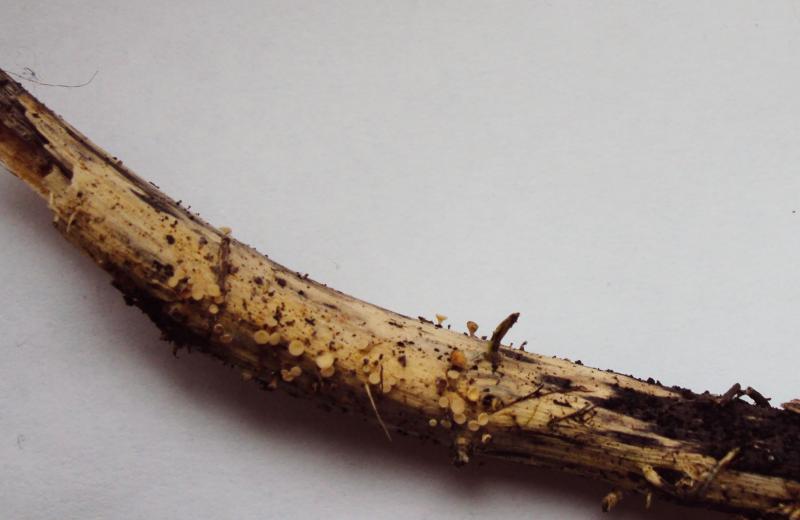
The 2d one was collected on a meadow, on rotten herbaceous stem.
Spores 9,1-12,7*1,8-2,7 um, fusiform, sometimes slightly S-shaped
Asci IKI B, with croziers, 44-60*3,8-5,5 um


in order to confuse you a bit :-)
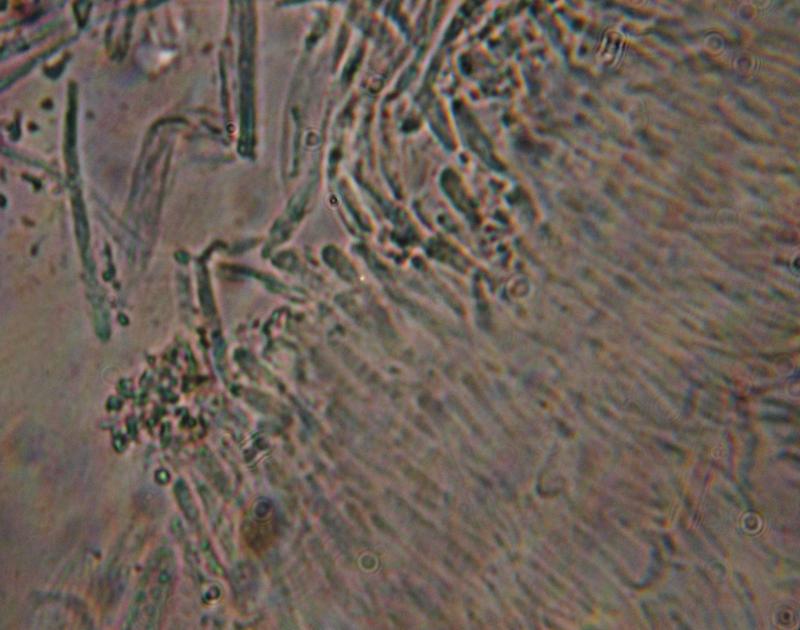
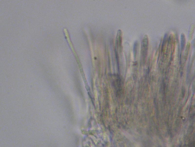
C. pallida is a species with marginal teeth, at least as I understand it. It was treated by Breitenbach & Kränzlin under the wrong name C. dolosella. The marginal teeth are not shown on their photo, but they are mentioned, and I reexamined their material:
your whitish specimen could well be C. cyathoidea, quite a variable species. Are the spores actually up to 3.6µm? Regrettably, only the spores are alive in your preparations. Maybe you press too strong. The apical ring photo seems to exclude hymenoscyphus repandus.
The brown one reminds me of C. cacaliae.
Zotto
Hello, Zotto!
And thank you for answer.
Yes, I know about marginal teeth in C. pallida, but in my opinion they probably could be poorly visible/destructed, so on. The 2d one was collected in dry condition, so I cannot say surely whether it was brown in living state or not.
With best regards,
Irina